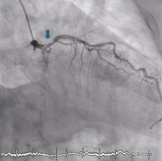





Delayed Intervention of a Thrombotic Saphenous Vein Graft Lesion
© 2026 HMP Global. All Rights Reserved.
Any views and opinions expressed are those of the author(s) and/or participants and do not necessarily reflect the views, policy, or position of the Journal of Invasive Cardiology or HMP Global, their employees, and affiliates.
J INVASIVE CARDIOL 2026. doi:10.25270/jic/26.00365. Epub October 21, 2026.
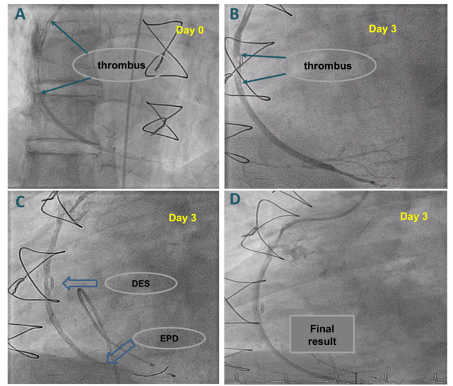
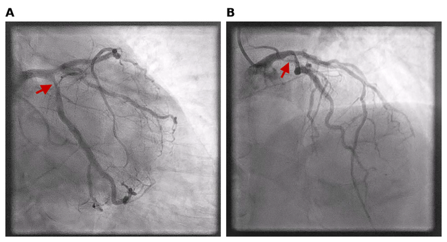
A 60-year-old man who had previously undergone coronary artery bypass graft surgery presented with non-ST-segment elevation myocardial infarction. Angiography showed chronic total occlusion of the left anterior descending (LAD) and right coronary artery, diffuse left circumflex disease, patent left internal mammary graft to the LAD, and patent saphenous vein graft (SVG) to the obtuse marginal branch. The culprit lesion was identified in the mid-segment of the SVG supplying the posterior descending artery, characterized by large thrombus burden and relatively impaired distal flow (Thrombolysis in Myocardial Infarction flow 2) (Figure A, Video 1).
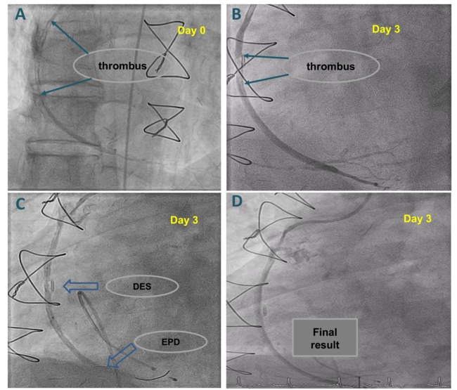
Immediate SVG percutaneous coronary intervention (PCI) was considered a high-risk procedure for distal embolization and subsequent no-reflow. Given that the patient was stable with no signs of ongoing ischemia, PCI was deferred and he was put on aspirin, ticagrelor, enoxaparin, and tirofiban for 72 hours in order to reduce the thrombotic burden of the SVG, thus facilitating PCI. Repeat angiography 3 days later showed marked thrombus reduction (Figure B, Video 2). Following the placement of a temporary pacemaker to treat bradycardia, PCI with the placement of a drug-eluting stent was performed. A FilterWire EZ Embolic Protection System (Boston Scientific) was advanced to the distal segment of the SVG to prevent distal thrombus embolism (Figure C, Video 3). Final angiography showed restored distal flow without residual stenosis (Figure D, Video 4).
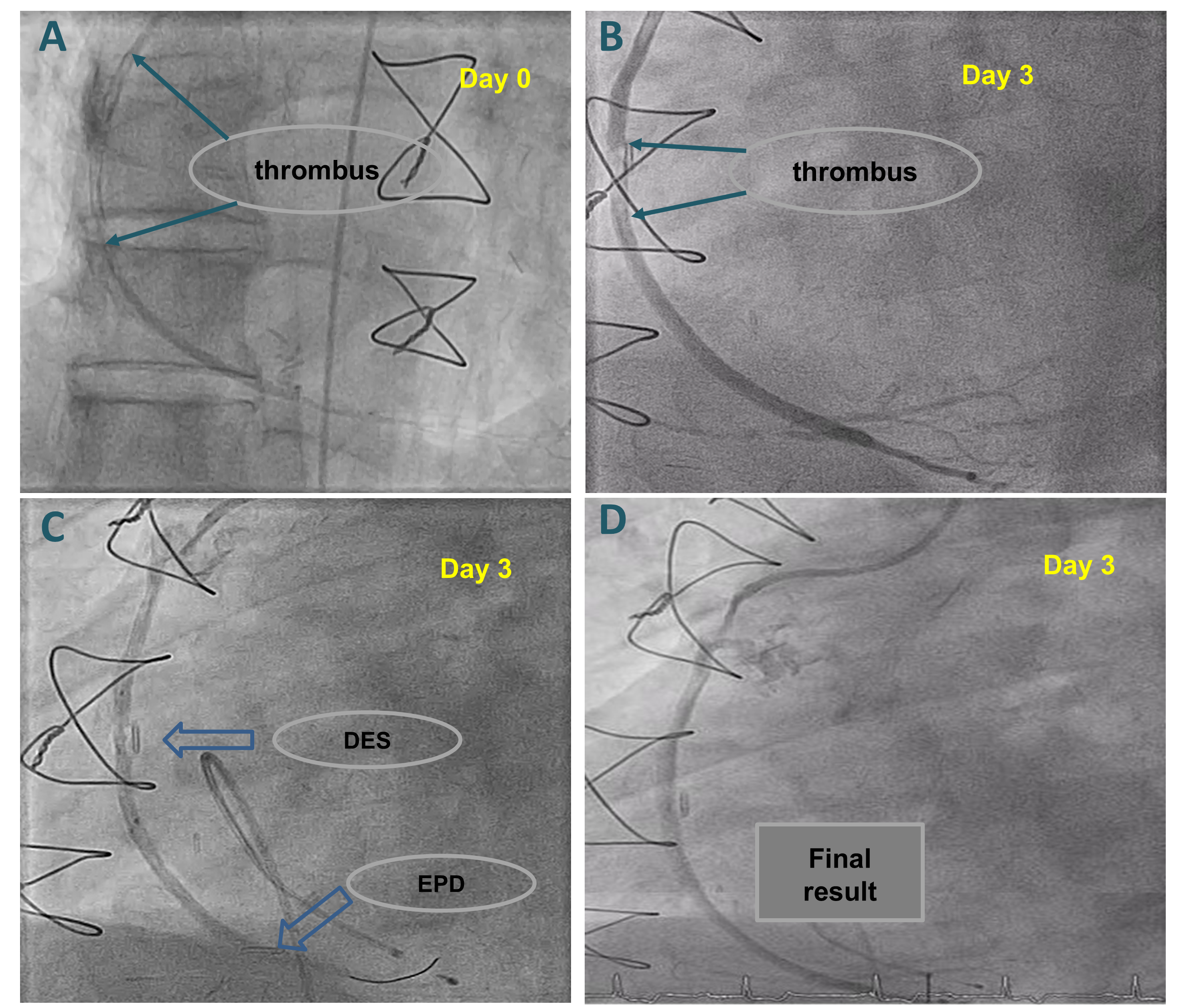
Recent data favor SVG PCI over corresponding coronary artery PCI.1 Nevertheless, SVG procedures are frequently complicated by large thrombus burden. Bulky, non-flow limiting thrombi in native coronary vessels have been initially treated successfully with a conservative approach with intense antithrombotic therapy.2 Our images illustrate that, in selected stable patients with SVG-related myocardial infarction, deferring PCI until after the administration of an intensive, short-term antithrombotic therapy regimen may facilitate intervention by drastically reducing the thrombus volume. A distal embolic protection device should be considered during SVG PCI when technically feasible.
Affiliations and Disclosures
Vasilki-Maria Dragona, MD1; Fotios Kolokathis, MD, PhD1; Emmanouil S. Brilakis, MD, PhD2; Iosif Xenogiannis, MD, PhD1
From the 1Second Department of Cardiology, Attikon University Hospital, National and Kapodistrian University of Athens, Chaidari, Athens, Greece; 2Minneapolis Heart Institute and Minneapolis Heart Institute Foundation, Abbott Northwestern Hospital, Minneapolis, Minnesota.
Disclosures: The authors report no financial relationships or conflicts of interest regarding the content herein.
Consent statement: The authors confirm that informed consent was obtained from the patient for the intervention described in the manuscript and to the publication including all images.
Address for correspondence: Iosif Xenogiannis, MD, PhD, Rimini 1, Chaidari, Athens 12461, Greece. Email: iosifxeno@hotmail.com
References
1. de Winter RW, Hoek R, Walsh SJ, et al; PROCTOR Trial Investigators. PCI of native coronary artery vs saphenous vein graft after prior bypass surgery: a multicenter, randomized trial. J Am Coll Cardiol. 2026;87(3):269-282. doi:10.1016/j.jacc.2025.09.1577
2. Xenogiannis I, Rallidis LS, Bakirtzis I, Pappas C, Κolokathis F. Large nonobstructive coronary thrombus in a patient with acute coronary syndrome: can it be treated conservatively? JACC Case Rep. 2025;30(6 Pt 1):102826. doi:10.1016/j.jaccas.2024.102826