




Capturing Coronary Artery Perforation on Optical Coherence Tomography: A Rare Imaging Record After Excimer Laser Atherectomy of a Calcified Nodule
© 2026 HMP Global. All Rights Reserved.
Any views and opinions expressed are those of the author(s) and/or participants and do not necessarily reflect the views, policy, or position of the Journal of Invasive Cardiology or HMP Global, their employees, and affiliates.
J INVASIVE CARDIOL 2026. doi:10.25270/jic/26.00366. Epub October 20, 2026.
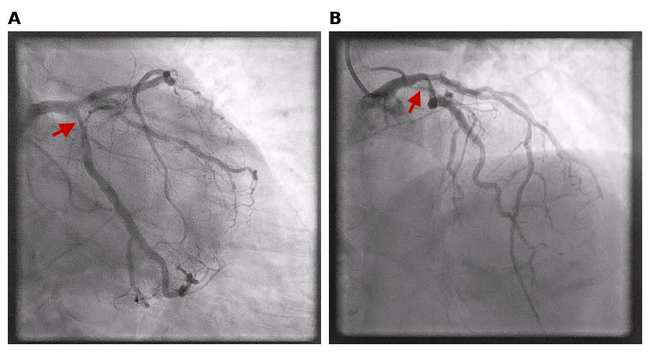
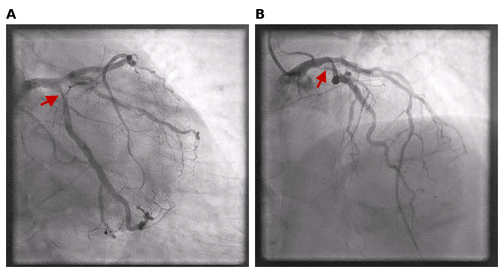
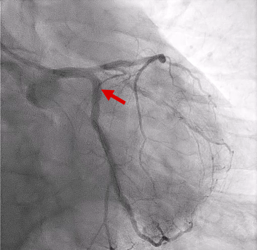
A 64-year-old man with chronic ischemic heart disease presented with non-ST-elevation myocardial infarction, Killip Class I. His history included prior ST-elevation myocardial infarction in 2021 treated with everolimus-eluting stents to the left anterior descending and mid-distal circumflex (LCx) arteries, and a 2024 admission for unstable angina, during which drug-coated balloon angioplasty was performed on the proximal LCx. Coronary angiography showed severe stenosis in the proximal LCx due to a calcified nodule, which was visible on 2 different projections (Figure 1).
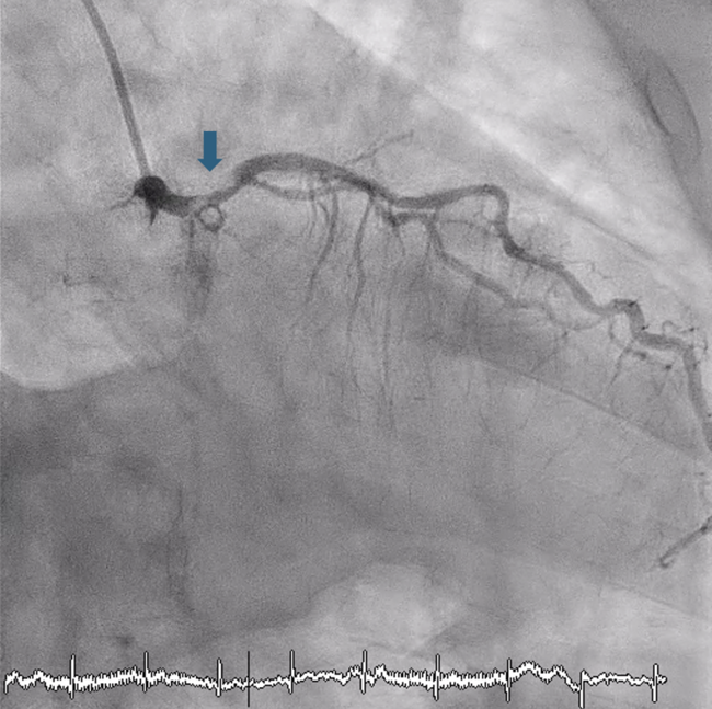
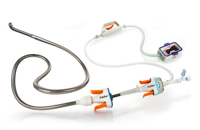
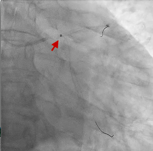
Percutaneous coronary intervention of the proximal LCx was performed via a 6F Extra Backup 3.5 Launcher guide catheter (Medtronic) and a Sion wire (Asahi Intecc) advanced to the distal LCx. An optical coherence tomography (OCT) catheter (Dragonfly OpStar, Abbott) could not be advanced through the lesion; thus, excimer laser plaque modification was performed with an ELCA coronary laser atherectomy catheter (Philips) (1.4 mm, 40 mJ/mm²/60 Hz, 1.4 min, 4000 pulses) (Figure 2).
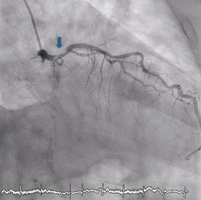
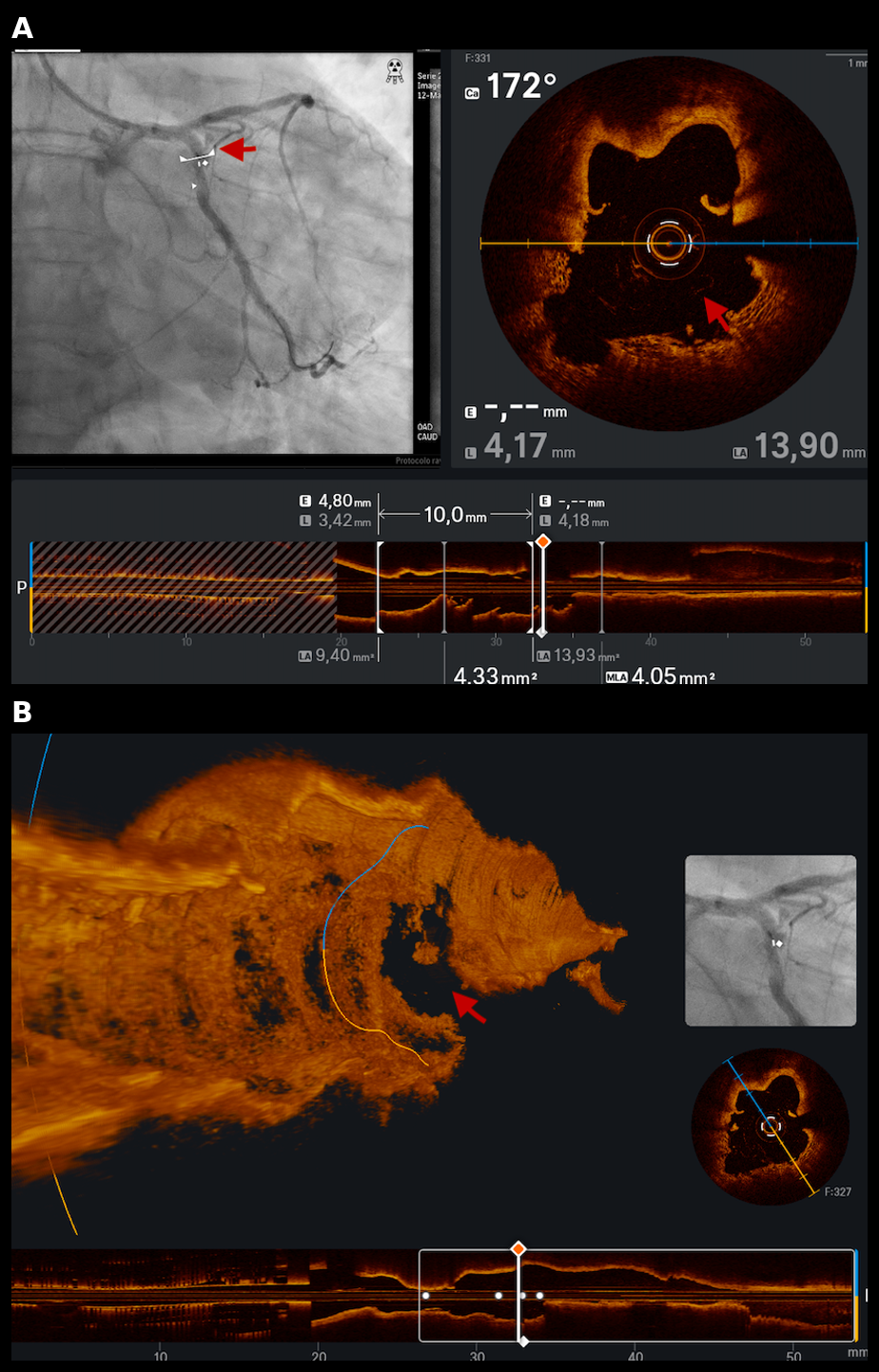
Post-laser OCT, together with angiography, revealed a coronary perforation with vessel wall rupture (Videos 1 and 2, Figure 3). Prolonged balloon inflation was performed with a 3.5 × 15-mm noncompliant (NC) TREK NEO balloon (Abbott). Given the OCT findings and to reduce the risk of pseudoaneurysm at follow-up, a 3.0 × 20-mm PK Papyrus covered stent (BIOTRONIK) was selected. Successful stent delivery required support from an Expressman guide extension catheter (APT Medical) combined with a balloon-anchoring technique. The stent was postdilated with a 3.5 × 15-mm NC TREK NEO balloon. Repeat OCT demonstrated an area of malapposition with persistent mild contrast leak, which resolved after further postdilation with a 4.0-mm NC TREK NEO balloon, achieving complete angiographic sealing of the perforation with an excellent final result (Figure 4).
The patient required transient low-dose vasopressor support for hypotension, and a minimal pericardial effusion was noted on echocardiography without evidence of tamponade. He was discharged without further complications and had no new events at the 5-month follow-up.
Affiliations and Disclosures
Oscar Lagos Degrande, MD; Alicia Cinta Argandoña Garrido, RN; Felix Rosa Longobardo, MD; Carmen Lluch Requerey, MD; Elena Izaga, PhD; Santiago Camacho Freire, PhD; Jessica Roa Garrido, MD; Antonio Gomez Menchero, MD
From the Department of Interventional Cardiology, Hospital Universitario Juan Ramón Jiménez, Huelva, Spain.
Disclosures: The authors report no financial relationships or conflicts of interest regarding the content herein.
Consent statement: The authors confirm that informed consent was obtained from the patient for the intervention(s) described in the manuscript and to the publication thereof, including any and all images.
Address for correspondence: Oscar Lagos Degrande, MD, Calle Opera Carmen 42, Seville 41007, Spain. Email: oldeg2450@gmail.com; X: @DegrandeLagosDr