




Clinical Videos
Video Supplement: Capturing Coronary Artery Perforation on Optical Coherence Tomography: A Rare Imaging Record After Excimer Laser Atherectomy of a Calcified Nodule
10/20/2026
Video Supplement to "Capturing Coronary Artery Perforation on Optical Coherence Tomography: A Rare Imaging Record After Excimer Laser Atherectomy of a Calcified Nodule" (Clinical Image).
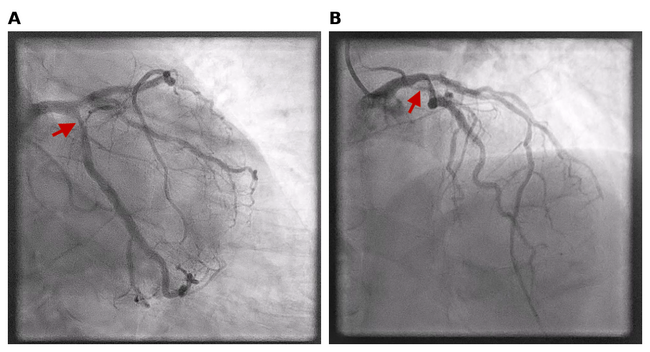
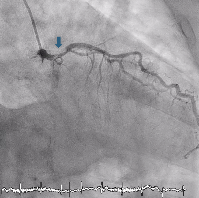
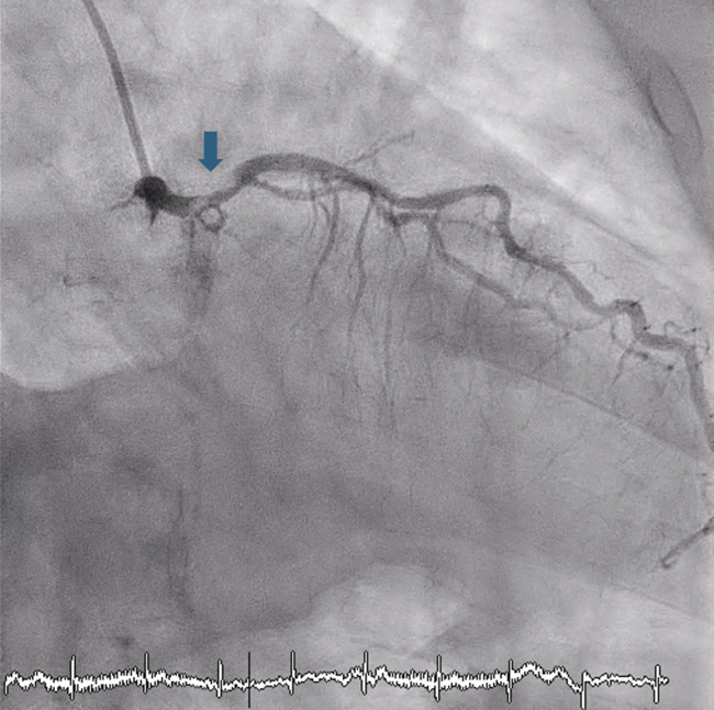
Video 1. Optical coherence tomography pullback through the proximal left circumflex artery after excimer laser atherectomy of a calcified nodule, showing a focal discontinuity of the vessel wall (red arrow), consistent with coronary artery perforation.
© 2026 HMP Global. All Rights Reserved.
Any views and opinions expressed are those of the author(s) and/or participants and do not necessarily reflect the views, policy, or position of the Journal of Invasive Cardiology or HMP Global, their employees, and affiliates.