




Impella 5.5–Facilitated Mitral Transcatheter Edge-to-Edge Repair in Cardiogenic Shock as a Bridge to Transplant Evaluation
© 2026 HMP Global. All Rights Reserved.
Any views and opinions expressed are those of the author(s) and/or participants and do not necessarily reflect the views, policy, or position of the Journal of Invasive Cardiology or HMP Global, their employees, and affiliates.
J INVASIVE CARDIOL 2026. doi:10.25270/jic/26.00339. Epub November 11, 2026.
Patients with severe functional mitral regurgitation (MR) and markedly enlarged coaptation gaps due to advanced dilated cardiomyopathy have largely been excluded from randomized trials evaluating mitral transcatheter edge-to-edge repair (M-TEER).1 Consequently, the role of M-TEER in patients with profound ventricular dilation and cardiogenic shock remains poorly defined. We present a case in which surgically implanted microaxial mechanical circulatory support with the Impella 5.5 (Abiomed) facilitated M-TEER in a patient with refractory cardiogenic shock and severe functional MR. Mechanical unloading with Impella support reduced the mitral coaptation gap, enabling successful transcatheter repair and clinical stabilization, allowing recovery from shock, early ambulation, and hospital discharge as a bridge to outpatient evaluation for orthotopic heart transplantation.
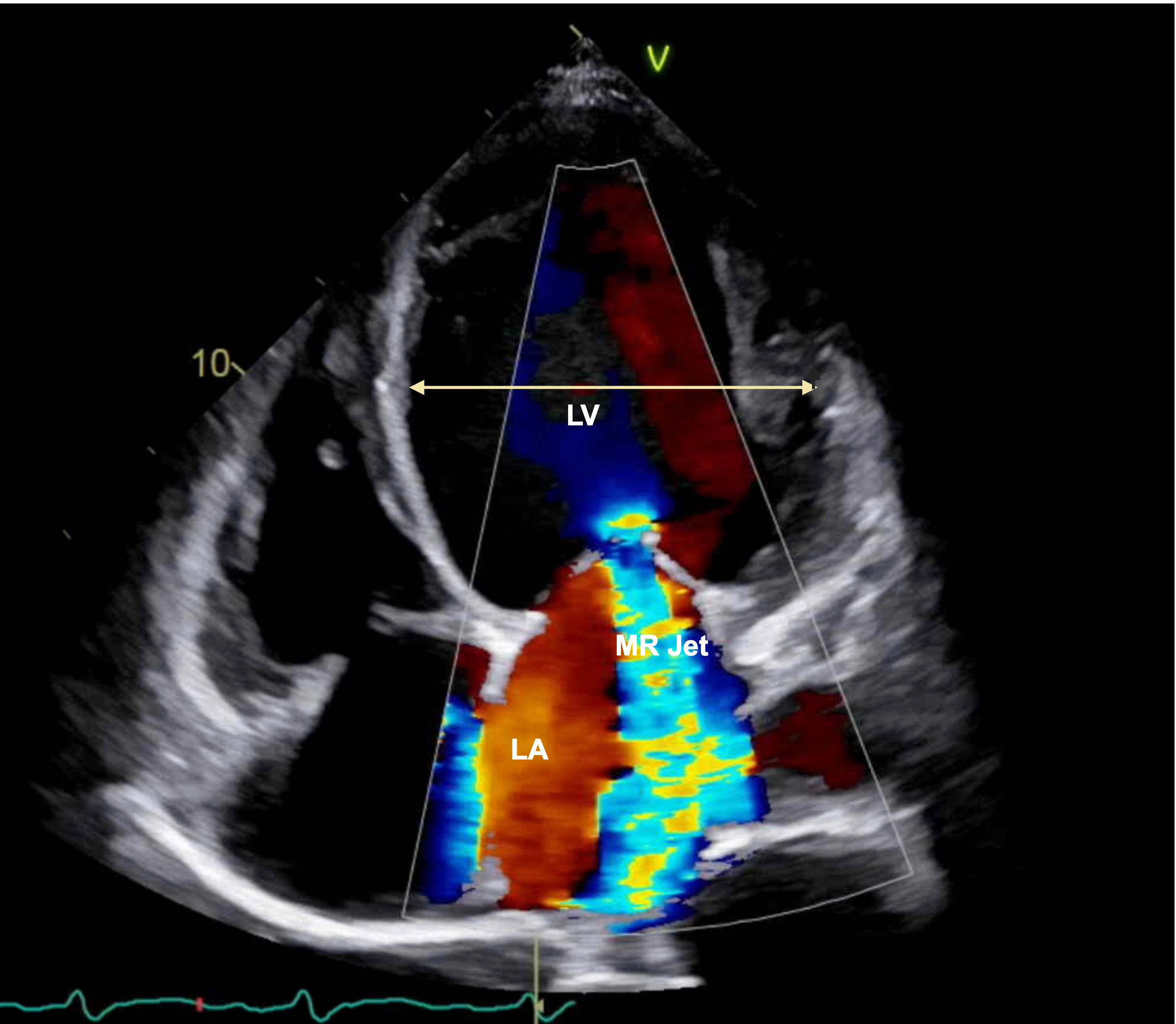
A 61-year-old woman with nonischemic cardiomyopathy presented with acute decompensated heart failure and progressive dyspnea. Transthoracic echocardiography demonstrated severely reduced left ventricular (LV) systolic function, marked ventricular dilation, and severe functional MR (Figure 1). Despite aggressive diuresis, transesophageal echocardiography confirmed persistent severe central MR due to non-coaptation of the anterior and posterior mitral leaflets (Figure 2).
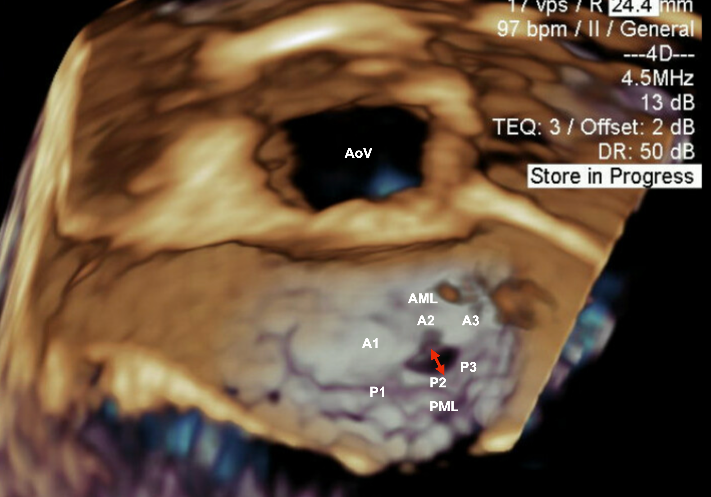
The patient subsequently developed flash pulmonary edema and cardiogenic shock requiring inotropic support with dobutamine and epinephrine, along with emergent placement of an Impella CP device. She was subsequently transitioned to surgically implanted Impella 5.5 support for ongoing mechanical circulatory assistance (Video).
Despite hemodynamic support and volume optimization, the patient remained dependent on Impella support and dobutamine, with worsening renal function and persistent severe MR on echocardiography (Video). Attempts to wean dobutamine were unsuccessful and resulted in worsening mixed venous oxygen saturation and clinical deterioration. Because heart transplantation and durable LV assist device services were not available at our institution, and insurance limitations precluded immediate transfer for transplant evaluation, a multidisciplinary heart team discussion was convened. Given the contribution of severe MR to ongoing hemodynamic instability, M-TEER was pursued.
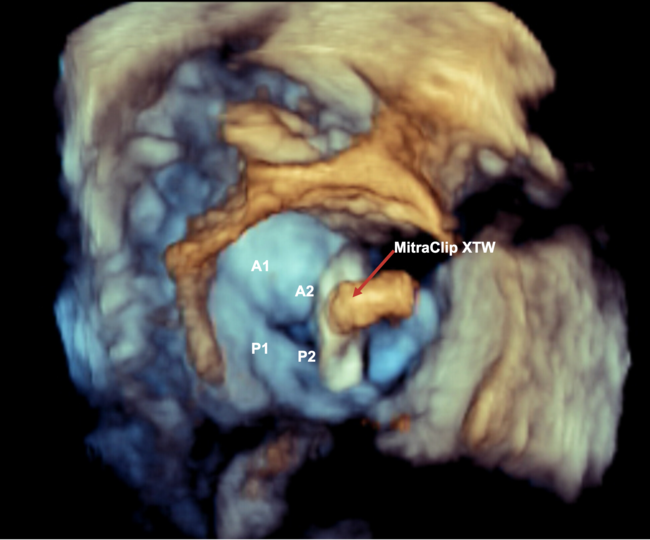
Impella-mediated LV unloading reduced the mitral coaptation gap, rendering M-TEER feasible (Figure 3). A MitraClip XTW device (Abbott) was used to approximate the A2–P2 scallops, resulting in a marked reduction in MR with immediate hemodynamic improvement. Mean left atrial pressure decreased by 10 mm Hg following clip deployment (Figure 4, Video).
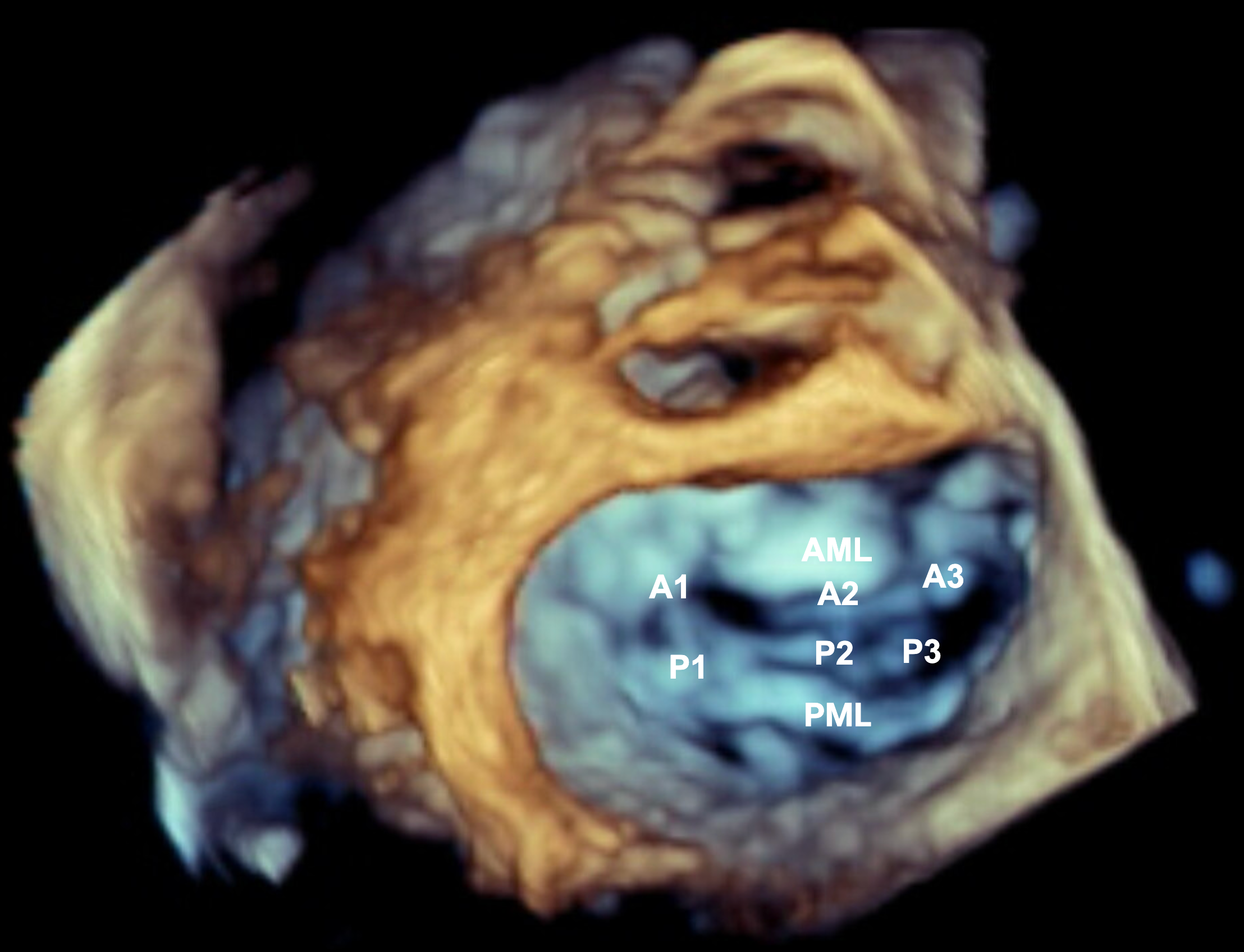
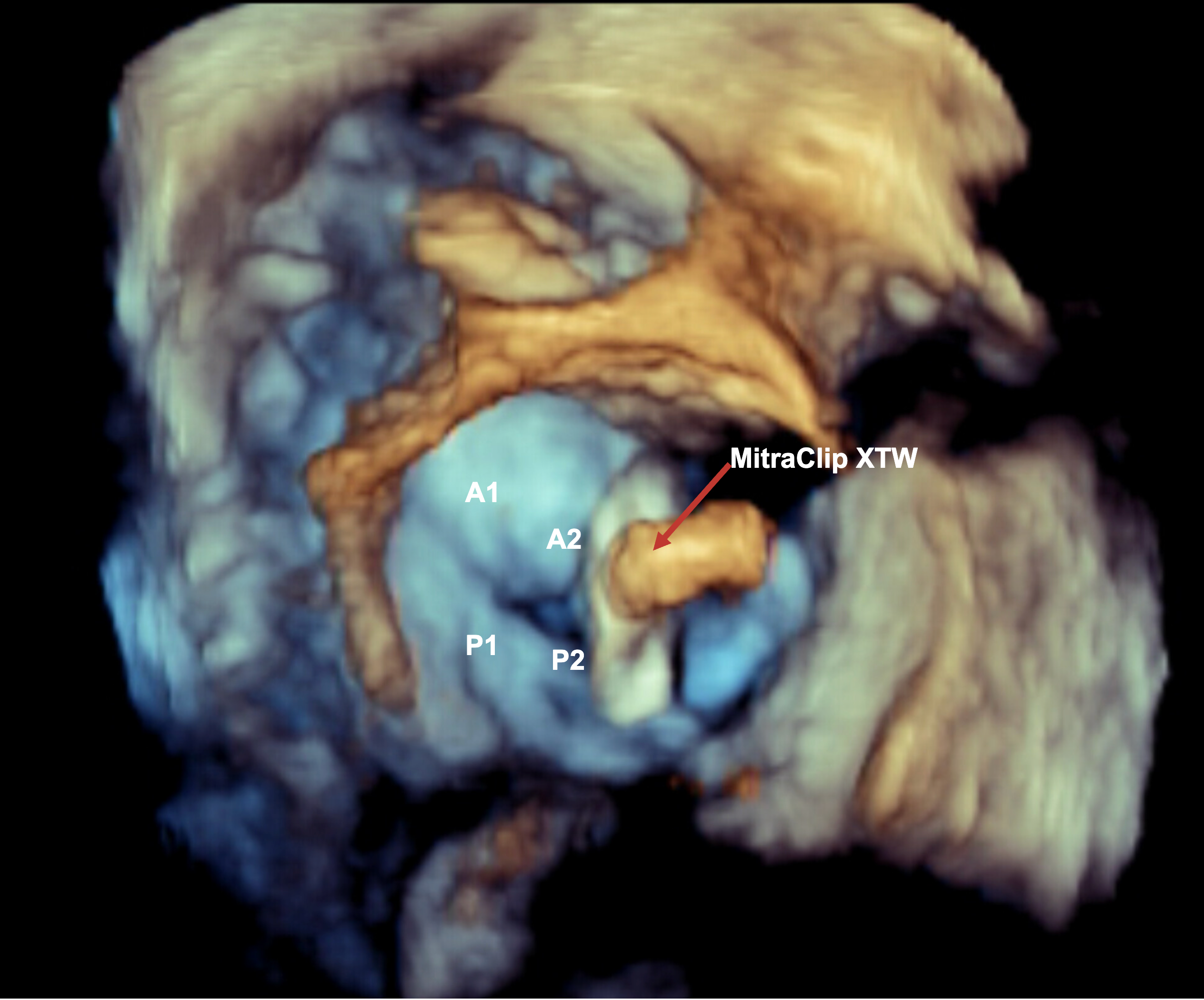
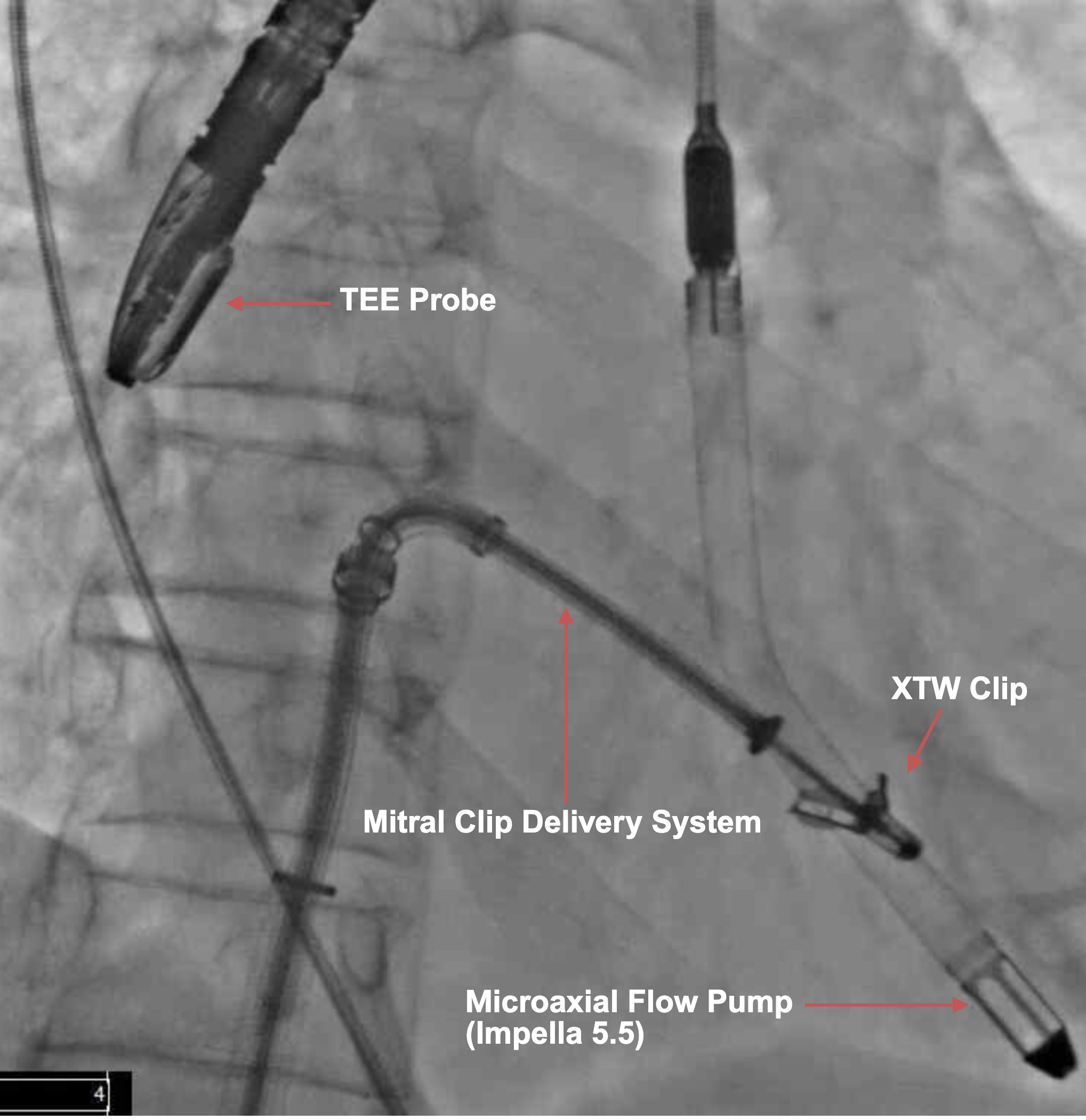
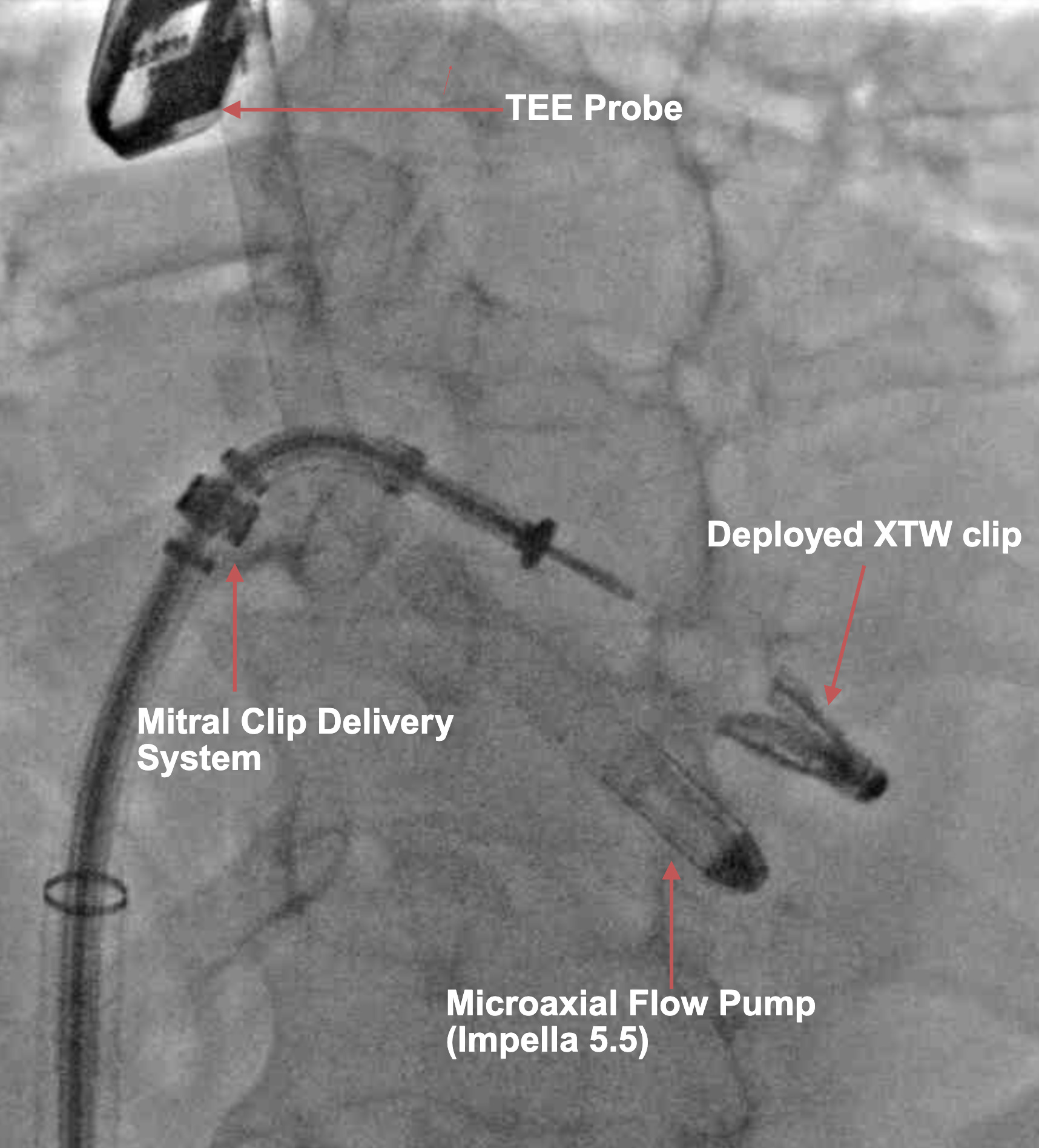
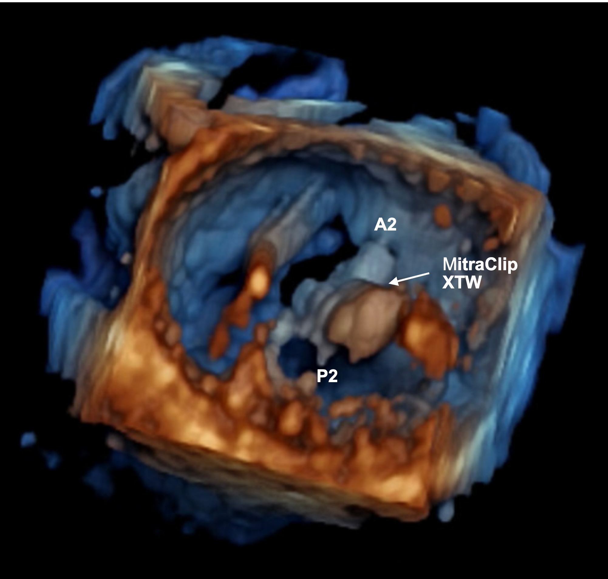
Over the following days, the patient demonstrated progressive hemodynamic improvement. Inotropic support was discontinued, and she was gradually mobilized while maintained on Impella 5.5 support. She subsequently underwent implantation of a cardiac resynchronization therapy–defibrillator. Renal function improved, allowing decannulation of the Impella 5.5 and hospital discharge on home milrinone therapy. At the 12-month follow-up, the patient was clinically stable without recurrent heart failure hospitalizations and undergoing evaluation for orthotopic heart transplantation.
This case highlights the role of Impella 5.5-mediated LV unloading to reduce the mitral coaptation gap and facilitate M-TEER in patients with severe functional MR and cardiogenic shock, suggesting a potential bridge to advanced heart failure therapies.
Affiliations and Disclosures
Arnav Kumar, MD, MSCR1; Hannan Chaugle, MD2
From the 1Structural Heart & Valve Center, Modern Heart & Vascular Institute, The University of Houston Tilman J. Fertitta Family College of Medicine, HCA Houston Healthcare, Houston, Texas; 2Department of Clinical Sciences, University of Houston College of Medicine, Surgical Specialists of Clear Lake, Houston, Texas.
Disclosures: The authors report no financial relationships or conflicts of interest regarding the content herein.
Consent statement: The authors confirm that informed consent was obtained from the patient(s) for the intervention(s) described in the manuscript and for the publication thereof, including any and all images.
Address for correspondence: Arnav Kumar, MD, MSCR, The University of Houston Tilman J. Fertitta Family College of Medicine, Modern Heart & Vascular Institute, HCA Houston Healthcare, 7400 Fannin St, Suite 650, Houston, TX 77054, USA. Email: structuralak@icloud.com; X: @arnavkumar
References
- Stone GW, Abraham WT, Lindenfeld J, et al; COAPT Investigators. Five-year follow-up after transcatheter repair of secondary mitral regurgitation. N Engl J Med. 2023;388(22):2037-2048. doi:10.1056/NEJMoa2300213