




Simplified Jailed Balloon Technique: Proof-of-Concept for Side Branch Preservation Without Routine Proximal Optimization or Final Kissing Balloon
© 2026 HMP Global. All Rights Reserved.
Any views and opinions expressed are those of the author(s) and/or participants and do not necessarily reflect the views, policy, or position of the Journal of Invasive Cardiology or HMP Global, their employees, and affiliates.
J INVASIVE CARDIOL 2026. doi:10.25270/jic/26.00268. Epub October 28, 2026.
Key Clinical Summary
- The Simplified Jailed Balloon Technique (S-JBT) prevents side branch (SB) occlusion during bifurcation stenting via simultaneous SB balloon inflation, without SB rewiring or final kissing balloon inflation.
- SB protection established by simultaneous inflation persisted after postdilation, indicating this initial step, not final optimization, is the primary mechanism preventing occlusion.
- Omitting rewiring and final kissing balloon inflation may shorten procedure time and contrast use for small SBs; proximal optimization remains appropriate when indicated.
Abstract
Objectives. The modified jailed balloon technique (M-JBT) decreases side branch (SB) occlusion during bifurcation percutaneous coronary intervention by simultaneously inflating an SB balloon alongside main vessel stent deployment. Conventionally, M-JBT requires subsequent proximal optimization (POT), SB rewiring, and final kissing balloon inflation (KBI), adding complexity to the procedure. The authors defined a simplified approach—the Simplified Jailed Balloon Technique (S-JBT)—where postdilation is performed using only the stent delivery balloon, omitting POT, rewiring, and final KBI.
Methods. To validate the physiological rationale of S-JBT, the authors measured SB resting full-cycle ratio (RFR) at 3 time points (baseline, post-M-JBT, and postdilation) in 6 consecutive patients undergoing S-JBT for LAD-diagonal bifurcation lesions using the jailed pressure wire technique.
Results. RFR was maintained at all timepoints (baseline: 0.82 ± 0.17; post-M-JBT: 0.84 ± 0.14; postdilation: 0.86 ± 0.15), with no angiographic SB compromise.
Conclusions. These findings suggest that the favorable SB ostial geometry established with simultaneous inflation persists after stent balloon postdilation, and that the primary mechanism of SB protection resides in the simultaneous inflation step. S-JBT may offer a practical, simplified SB protection strategy, particularly for bifurcation lesions with small side branches.
Introduction
In percutaneous coronary intervention (PCI) for coronary bifurcation lesions, side branch (SB) occlusion occurs in 5% to 7% of cases and is associated with an increased risk of myocardial infarction and cardiac death.1 The jailed wire technique (JWT), while straightforward, may provide insufficient protection in high-risk lesions. In contrast, the modified jailed balloon technique (M-JBT) substantially reduces SB occlusion by simultaneously inflating an SB balloon—positioned with its proximal marker at the SB ostium—alongside main vessel (MV) stent deployment.2
However, M-JBT typically requires proximal optimization (POT), SB rewiring, and final kissing balloon inflation (KBI), adding procedural complexity and operator burden. Moreover, the clinical benefit of routine KBI has been increasingly questioned, with several trials demonstrating no consistent improvement in outcomes.3 In routine clinical practice, we repeatedly observed preserved SB flow after M-JBT followed by postdilation with the stent delivery balloon alone, without POT or KBI. Here, we describe this simplified approach, the Simplified Jailed Balloon Technique (S-JBT), and aim to validate its physiological rationale using the JWT.
Methods
A conceptual concern with S-JBT is whether postdilation with the stent delivery balloon removes the benefit of simultaneous inflation, rendering the technique functionally equivalent to JWT. To address this question, we measured SB resting full-cycle ratio (RFR) at 3 time points— (1) baseline, (2) immediately after M-JBT, and (3) after stent balloon postdilation—in 6 consecutive patients undergoing S-JBT for left anterior descending artery (LAD)-diagonal bifurcation lesions using JWT.
The study was conducted following institutional guidelines and the tenets of the Declaration of Helsinki and was approved by the ethics committee of Chubu Tokushukai Hospital (approval no. 26-14). The authors confirm that informed consent was obtained from the patients for the study and interventions described in the manuscript, and for the publication of their data, including any and all images.
S-JBT: definition and procedure
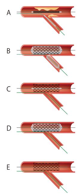
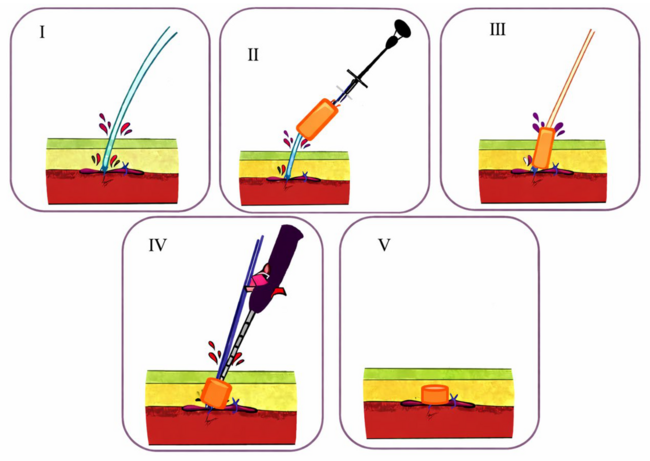
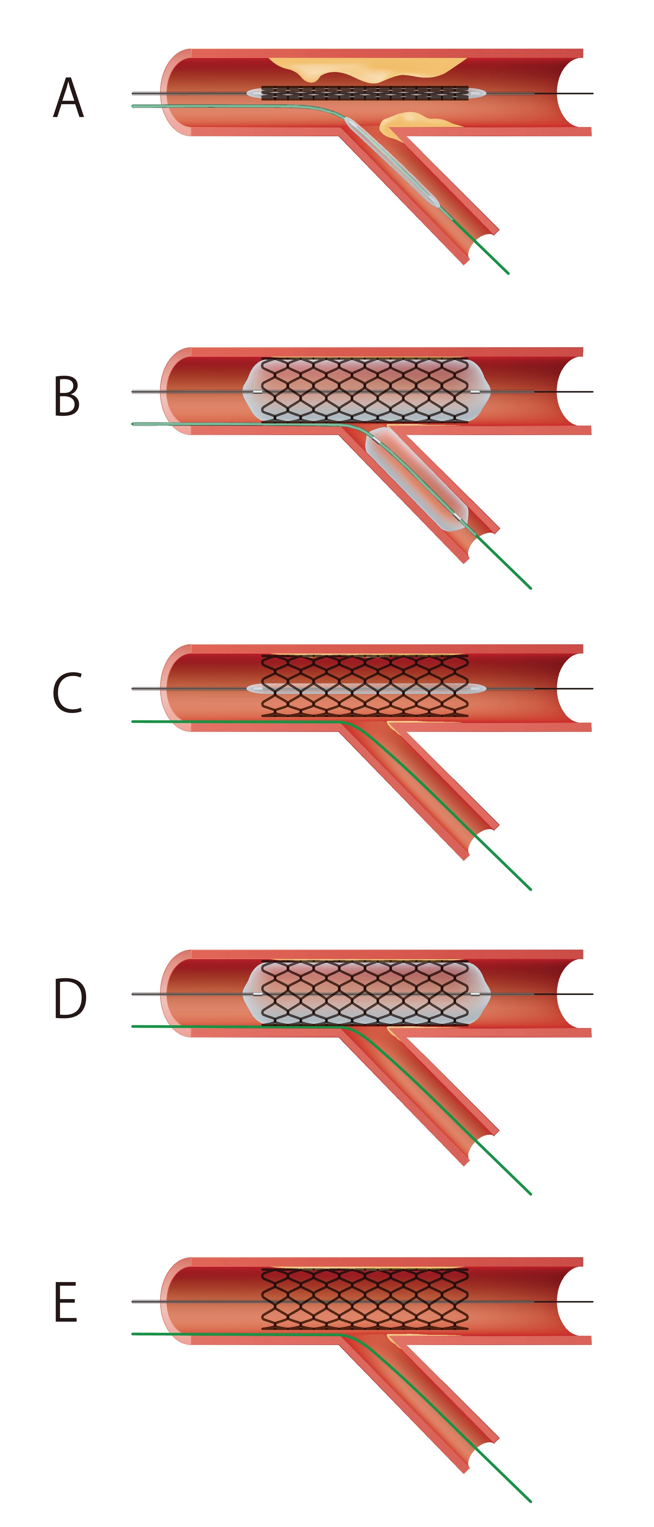
The procedural schema of S-JBT is illustrated in Figure 1. Guidewires are advanced into both the MV and SB. An SB protection balloon is positioned with its proximal marker at the SB ostium, and the MV stent is simultaneously deployed (M-JBT step). Then, the SB balloon is withdrawn while the SB wire remains jailed. Postdilation is performed using only the stent delivery balloon. POT, SB rewiring, and final KBI are intentionally omitted.
Representative case
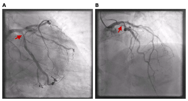
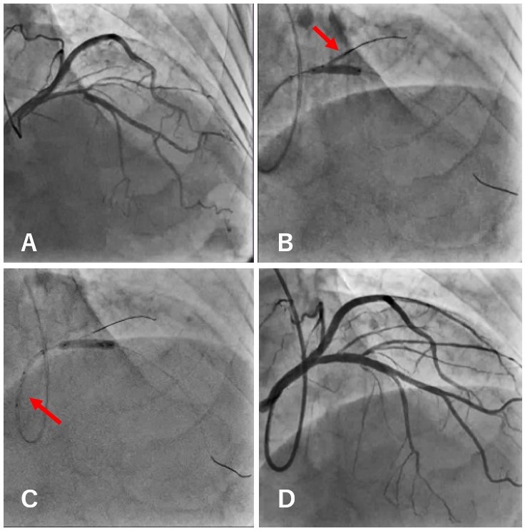
A 64-year-old man with hypertension and a history of smoking presented with exertional chest pain. Coronary angiography revealed 75% stenosis at the proximal LAD, segment 6 (Medina classification 1, 0, 0). After predilation with a 3.75 x 15-mm noncompliant balloon, angiography demonstrated 99% flow delay in the first diagonal branch (D1), requiring aggressive SB protection. S-JBT was performed using a 1.75 x 15-mm semicompliant balloon in D1 and a 4.0 x 18-mm drug-eluting stent in the MV. Final angiography confirmed preserved D1 flow (Figure 2).
Results
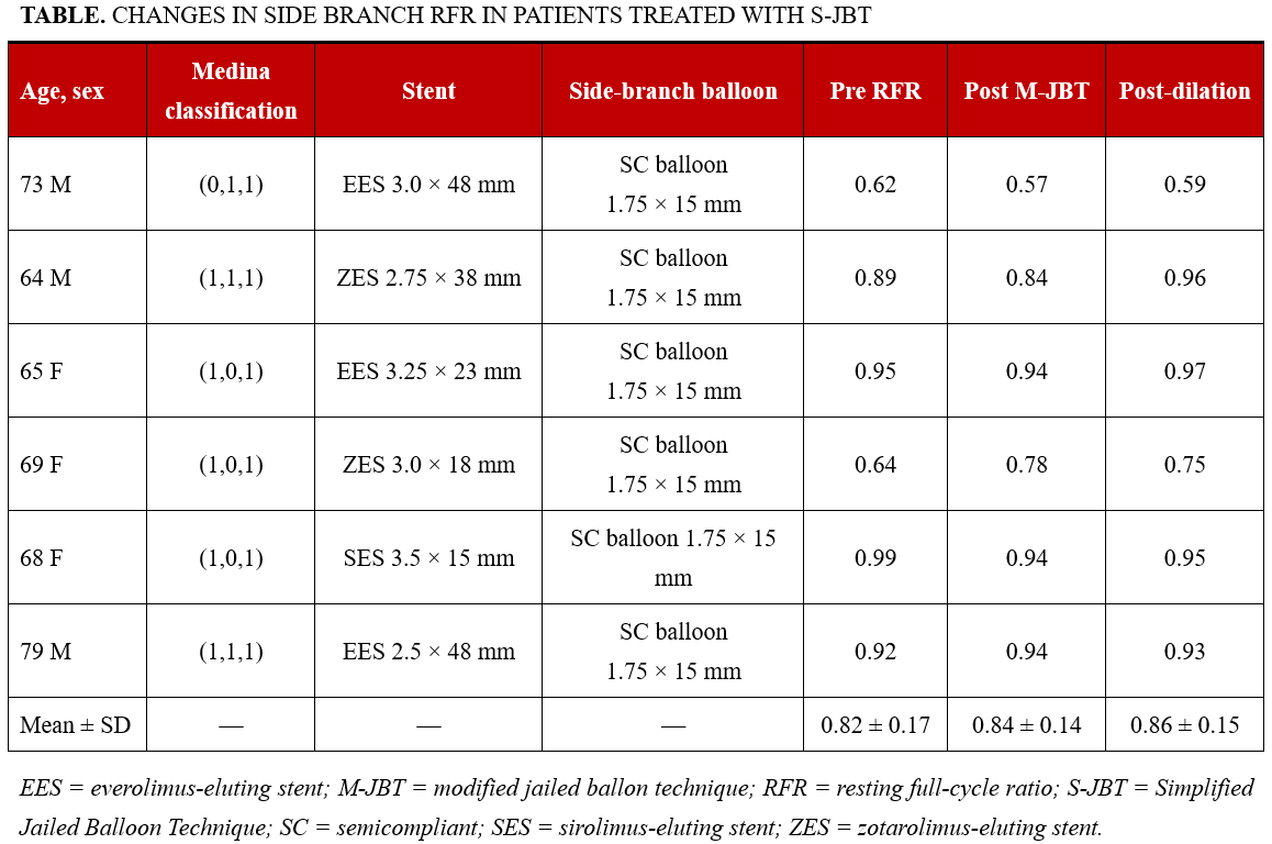
SB flow was angiographically preserved in all 6 cases, with no acute occlusion or slow flow. RFR values are summarized in the Table. Mean RFR was 0.82 ± 0.17 at baseline, 0.84 ± 0.14 after M-JBT, and 0.86 ± 0.15 after postdilation, with no deterioration at any time point. None of the cases demonstrated a marked RFR decrease after postdilation.
Discussion
Unlike JWT, where carina shift and plaque redistribution may compromise SB during stenting, M-JBT preserves SB ostial geometry through simultaneous balloon inflation. These findings suggest that the favorable ostial geometry established at stenting is maintained even after subsequent postdilation with the stent delivery balloon. The intentional POT and KBI omission did not impair SB physiology, supporting the hypothesis that the primary mechanism of SB protection in M-JBT arises from the simultaneous inflation itself rather than from final optimization maneuvers. This is consistent with prior randomized trials showing no consistent prognostic benefit of routine KBI.3
Qiu et al recently reported an active jailed balloon technique (A-JBT). 4 In A-JBT, the SB balloon is inflated at a higher pressure before the MV stent balloon, resulting in favorable angiographic outcomes. Although A-JBT and S-JBT share balloon-based SB protection, the two differ in their balloon inflation sequence and pressure settings. In S-JBT, as in M-JBT, the SB balloon and MV stent balloon are inflated simultaneously to nominal pressure. Therefore, our findings should not be considered a physiological validation of A-JBT. Our study’s serial RFR assessment offers physiological insights into the sequence of M-JBT.
Regarding POT, we acknowledge its established role in bifurcation PCI.5 Its omission in the present series was intentional in order to isolate the protective effect of S-JBT and should not be interpreted as a denial of its utility. POT remains appropriate when stent underexpansion or malapposition is suspected. Eliminating rewiring and KBI offers other practical benefits, such as shorter procedure time, reduced contrast use, and the avoidance of the technical difficulty and stent deformation risk associated with SB recrossing.
Limitations
Limitations include the small sample size (6 cases), the absence of a control group, the single-center retrospective design, and the restriction to LAD-diagonal bifurcations. Applicability to left main bifurcation lesions and other anatomies remains to be established. Prospective studies are warranted to confirm S-JBT efficacy and safety.
Conclusions
S-JBT is a simplified bifurcation PCI strategy that maintains SB physiological function after postdilation and does not require POT, SB rewiring, or final KBI. This approach may represent a practical SB protection strategy, particularly for bifurcation lesions with small SBs.
Affiliations and Disclosures
Jumpei Todoroki, MD; Ryuji Shiraishi, MD; Tasuku Sakurai, MD; Kenichiro Higa, MD
From the Department of Cardiology, Chubu Tokushukai Hospital, Kitanakagusuku, Japan.
Disclosures: The authors report no financial relationships or conflicts of interest regarding the content herein.
Address for correspondence: Jumpei Todoroki, MD, Department of Cardiology, Chubu Tokushukai Hospital, 801 Higa, Kitanakagusuku-son, Nakagami-gun, Okinawa 901-2393, Japan. Email: pureandpeace.go@gmail.com
References
1. Lassen JF, Holm NR, Banning A, et al. Percutaneous coronary intervention for coronary bifurcation disease: 11th consensus document from the European Bifurcation Club. EuroIntervention. 2016;12(1):38-46. doi:10.4244/EIJV12I1A7
2. Saito S, Shishido K, Moriyama N, et al. Modified jailed balloon technique for bifurcation lesions. Catheter Cardiovasc Interv. 2018;92(3):E218-E226. doi:10.1002/ccd.27334
3. Kim YH, Lee JH, Roh JH, et al. Randomized comparisons between different stenting approaches for bifurcation coronary lesions with or without side branch stenosis. JACC Cardiovasc Interv. 2015;8(4):550-560. doi:10.1016/j.jcin.2015.01.016
4. Qiu J, Li L, Wang W, et al. Jailed high-pressure balloon technique is superior to jailed wire technique in protecting side branch of coronary bifurcation lesions. Scand Cardiovasc J. 2024;58(1):2347295. doi:10.1080/14017431.2024.2347295
5. Lassen JF, Burzotta F, Banning AP, et al. Percutaneous coronary intervention for the left main stem and other bifurcation lesions: 12th consensus document from the European Bifurcation Club. EuroIntervention. 2018;13(13):1540-1553. doi:10.4244/EIJ-D-17-00622