




Reestablishing Aortic Continuity: Transcatheter Repair of Interrupted Aortic Arch
© 2026 HMP Global. All Rights Reserved.
Any views and opinions expressed are those of the author(s) and/or participants and do not necessarily reflect the views, policy, or position of the Journal of Invasive Cardiology or HMP Global, their employees, and affiliates.
J INVASIVE CARDIOL 2026. doi:10.25270/jic/26.00380. Epub November 16, 2026.
A 25-year-old man with a 4-year history of systemic hypertension presented with a new-onset severe headache. He had a history of subarachnoid hemorrhage requiring surgical clipping of an intracranial aneurysm in the middle cerebral artery territory 4 years prior. Clinical examination revealed a marked blood pressure discrepancy between the right upper limb (170/80 mm Hg) and the right lower limb (90/78 mm Hg), strongly suggesting significant aortic coarctation.
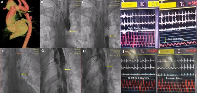
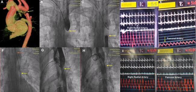
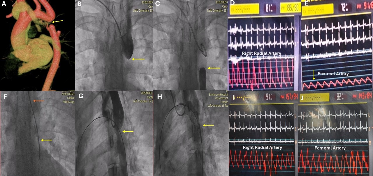
Transthoracic echocardiography demonstrated a narrowed aortic segment immediately distal to the left subclavian artery, with a characteristic Doppler waveform showing late systolic peaking and a persistent diastolic tail. Left ventricular systolic function was preserved, with an ejection fraction of 55% to 60%; no significant valvular abnormalities were identified. The patient underwent a computed tomography aortogram, which revealed complete interruption of the aorta just distal to the left subclavian artery (Figure A).
We offered surgical correction; however, given the surgical risks, the patient opted for an endovascular approach. We planned a catheter-based intervention using femoral and bilateral radial arterial access. Angiographic assessment and invasive pressure measurements helped in procedural planning. The invasive aortogram revealed a blunt stump just distal to the subclavian takeoff and another blunt stump distally (Figure B and C, Videos 1 and 2). The pressure gradient across the segment was approximately 100 mm Hg (Figure D and E). A complex wire-and-catheter technique, incorporating contralateral access, wire externalization, and snare assistance, enabled secure traversal of the coarctation segment (Figure F). Initial balloon dilatation was performed with a 10 × 30-mm balloon, followed by deployment of a 34-mm Zephyr XL stent (Sahajanand Laser Technology Ltd), with further expansion using a 16 × 40-mm Atlas Gold balloon (BD) (Figure G and H, Video 3). There was a residual gradient of only 10 mm Hg at the end of the procedure (Figure I and J).
Interrupted aortic arch (IAA) is a rare congenital aortic malformation, accounting for less than 1% of congenital heart defects. It is characterized by complete anatomical discontinuity between the ascending and descending aorta and represents the most severe form of aortic arch obstruction. IAA is classified into 3 subtypes according to the site of interruption: type A, distal to the origin of the left subclavian artery (LSA); type B, between the left common carotid artery (LCCA) and the LSA; and type C, between the innominate artery and the LCCA. Type B is the most common subtype in neonates, accounting for about 53% of cases, whereas type A predominates in adults, making up about 79% of reported cases.1 The choice of surgical technique depends on the anatomical characteristics and site of the aortic interruption. In adults, extra-anatomical bypass conduit repair performed as a single-stage procedure is the most commonly employed surgical approach. Although technically challenging, percutaneous stent implantation is a feasible and effective alternative to conventional surgical repair in appropriately selected patients.2
This case highlights the importance of recognizing aortic coarctation in young adults presenting with persistent hypertension and marked upper-to-lower limb blood pressure gradients. Endovascular stenting is a valuable therapeutic strategy to relieve obstruction and potentially improve long-term blood pressure control.
Affiliations and Disclosures
Devesh Kumar, MD, MRCP, DM1; Ranjit Nath, MD, DM, FACC, FESC1; Anwar Hussan Ansari, MD, DM1; Suad Akhtar, MD, DM1; Sonu Pandit, MD1; Shrividya Rao, MS, MCh2; Pahul Ahuja, MD1; Puneet Gupta, MD, DM1; Anunay Gupta, MD, DM1
From the Departments of 1Cardiology and 2Cardiothoracic & Vascular Surgery, Vardhman Mahavir Medical College and Safdarjung Hospital, New Delhi, India.
Disclosures: The authors report no financial relationships or conflicts of interest regarding the content herein.
Consent statement: The authors confirm that they obtained informed consent from the patient for the intervention described in the manuscript and for its publication, including images.
Address for correspondence: Ranjit Nath, Department of Cardiology, Vardhman Mahavir Medical College and Safdarjung Hospital, New Delhi 110029, India. Email: ranjithnath@yahoo.com
References
1. Erden I, Kayapinar O, Erden EC, Yalçin S. Silent interrupted aortic arch in an elderly patient. Cardiol J. 2011;18(6):695-697. doi:10.5603/cj.2011.0036
2. Kumar D, Ansari AH, Malik A, et al. Transcatheter stenting for severe native aortic coarctation. J Invasive Cardiol. 2026. doi:10.25270/jic/26.00265