Giant Hepatic Artery Aneurysm Presenting With a Hyperattenuating Crescent Sign: Successful Single-Plug Endovascular Exclusion
© 2026 HMP Global. All Rights Reserved.
Any views and opinions expressed are those of the author(s) and/or participants and do not necessarily reflect the views, policy, or position of the Journal of Invasive Cardiology or HMP Global, their employees, and affiliates.
J INVASIVE CARDIOL 2026. doi:10.25270/jic/26.00276. Epub August 21, 2026.
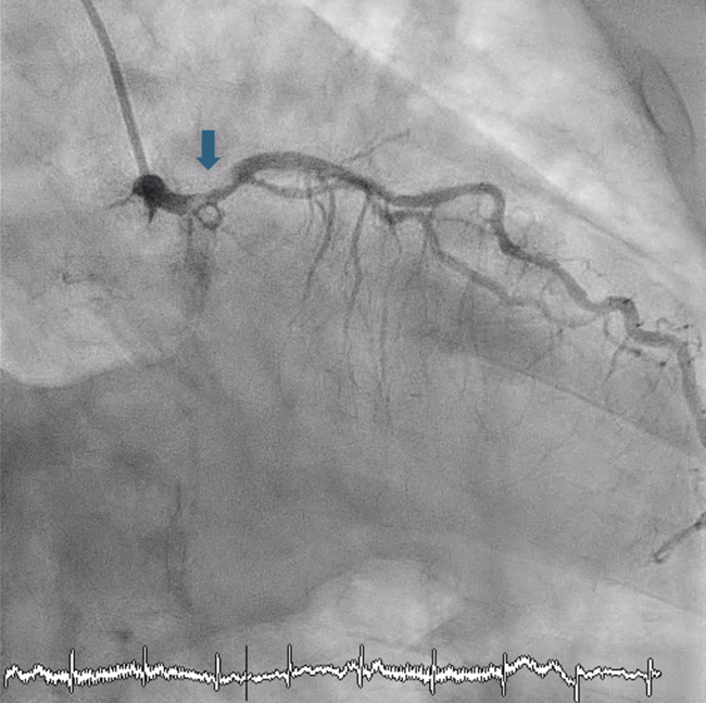
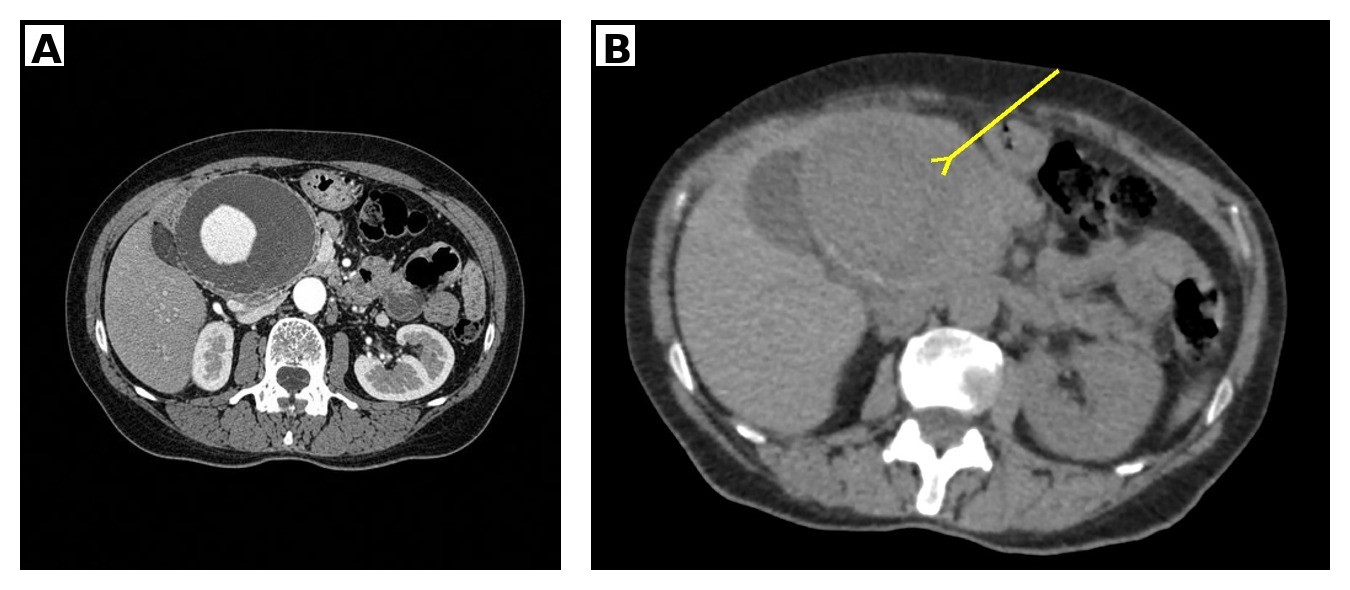
A 65-year-old woman with a history of smoking presented with a 30-day history of epigastric pain, nausea, and asthenia, with acute worsening on the day of admission. Contrast-enhanced computed tomography (CT) demonstrated a giant, fusiform aneurysm of the common hepatic artery measuring 7.5 × 7.5 cm with extensive mural thrombus (Figure 1A) and a hyperattenuating crescent sign (Figure 1B, arrow), indicating intramural hemorrhage and impending rupture. The aneurysm exerted mass effect on the portal vein, inferior vena cava, and right renal artery (Figure 1C), with preserved hepatopetal portal venous flow on venous-phase imaging.
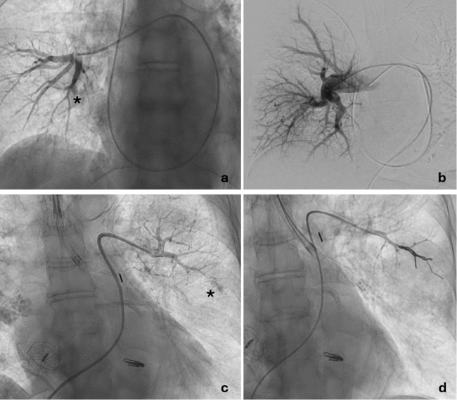
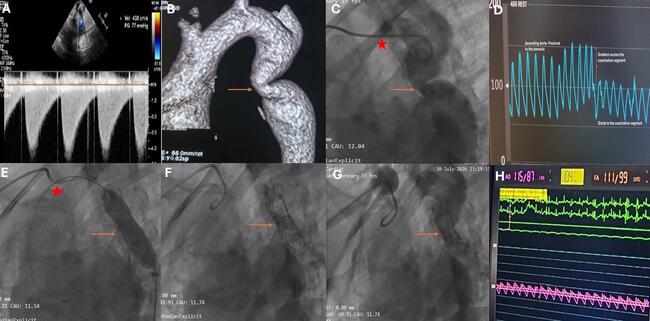
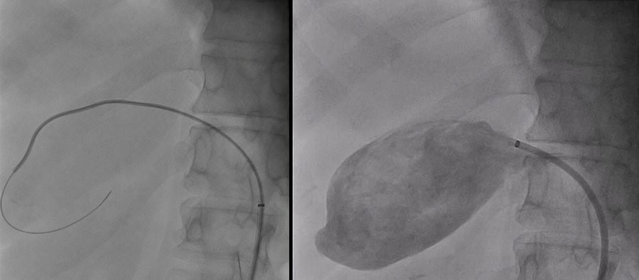
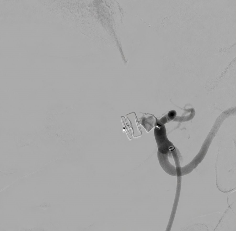
Selective celiac angiography confirmed the aneurysm, with complete absence of antegrade opacification of the distal hepatic arterial branches, consistent with spontaneous distal thrombosis (Video 1, Figure 2A). After multidisciplinary discussion, a single 14-mm Amplatzer Vascular Plug II (Abbott) was deployed at the aneurysm neck through a 6F guiding sheath using a telescoping technique that preserved the left gastric artery origin (Figure 2B, arrow; Video 2). Completion angiography confirmed complete interruption of aneurysmal inflow (Figure 3, Video 3).
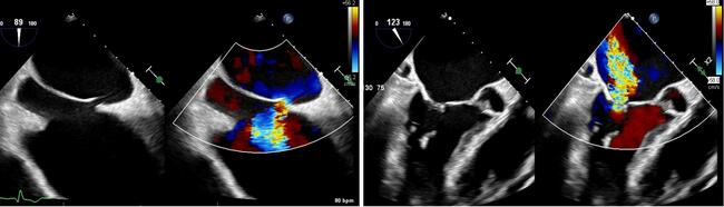
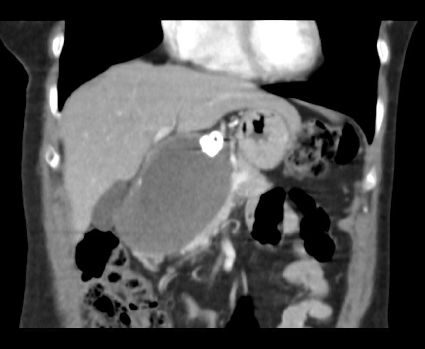
No hepatic ischemia occurred post-procedure. At the 4-month follow-up, CT confirmed persistent aneurysm exclusion without recanalization or hepatic parenchymal injury and with partial regression in aneurysm sac size and reduction of mass effect on adjacent structures, and the plug in stable position (Figure 4).
This case illustrates the hyperattenuating crescent sign as a marker of impending rupture in a giant visceral artery aneurysm and shows that angiographically confirmed absence of distal arterial inflow can allow isolated proximal plug occlusion, simplifying endovascular treatment of an otherwise high-risk lesion.
Affiliations and Disclosures
Ricardo de Souza Alves Ferreira, MD, MSc1; Guilherme Evangelista Rezende, MD1; Raphael França Lacerda de Andrade, MD2; José Luís Attab dos Santos, MD1,2
From the 1Hemodynamics and Interventional Cardiology Service (HCI-HRCor), Santa Casa de Misericórdia de São Sebastião do Paraíso, Minas Gerais, Brazil; 2Hemodynamics and Interventional Cardiology Service, HCI – Santa Casa de Ribeirão Preto, Ribeirão Preto, São Paulo, Brazil.
Disclosures: The authors report no financial relationships or conflicts of interest regarding the content herein.
Consent statement: The authors confirm that informed consent was obtained from the patient for the intervention(s) described in the manuscript and to the publication thereof, including any and all images.
Artificial intelligence (AI) statement: An AI-assisted tool (Claude, Anthropic) was used to assist with drafting and condensing the manuscript text to meet journal word-count requirements; the authors reviewed, edited, and verified all clinical statements. No figures, images, or other visual content were generated or modified using AI. All figures are original, unmodified clinical computed tomography and angiographic images provided directly by the authors and composed using standard image-editing software.
Address for correspondence: Ricardo de Souza Alves Ferreira, MD, MSc, Hemodynamics and Interventional Cardiology Service (HCI-HRCor), Santa Casa de Misericórdia de São Sebastião do Paraíso, Minas Gerais 37950-000, Brazil. Email: ricardoferreira@doctor.com
References
1. Abbas MA, Fowl RJ, Stone WM, et al. Hepatic artery aneurysm: factors that predict complications. J Vasc Surg. 2003;38(1):41-45. doi:10.1016/s0741-5214(03)00090-9
2. Chaer RA, Abularrage CJ, Coleman DM, et al. The Society for Vascular Surgery clinical practice guidelines on the management of visceral aneurysms. J Vasc Surg. 2020;72(1S):3S-39S. doi:10.1016/j.jvs.2020.01.039
3. Mehard WB, Heiken JP, Sicard GA. High-attenuating crescent in abdominal aortic aneurysm wall at CT: a sign of acute or impending rupture. Radiology. 1994;192(2):359-362. doi:10.1148/radiology.192.2.8029397
4. Van Holsbeeck A, Dhondt E, Marrannes J, et al. Coil embolization of true, common and proper, hepatic artery aneurysms: technique, safety and outcome. Cardiovasc Intervent Radiol. 2023;46(4):480-487. doi:10.1007/s00270-023-03379-0
5. Tuite DJ, Kessel DO, Nicholson AA, Patel JV, McPherson SJ, Shaw DR. Initial clinical experience using the Amplatzer Vascular Plug. Cardiovasc Intervent Radiol. 2007;30(4):650-654. doi:10.1007/s00270-007-9044-3