Recurrent Acute Pericarditis in a Patient With History of Acute Rheumatic Fever: The Role of Cardiac Magnetic Resonance in Diagnosis and Management
© 2026 HMP Global. All Rights Reserved.
Any views and opinions expressed are those of the author(s) and/or participants and do not necessarily reflect the views, policy, or position of the Journal of Invasive Cardiology or HMP Global, their employees, and affiliates.
J INVASIVE CARDIOL 2026. doi:10.25270/jic/26.00259. Epub August 13, 2026.
A 23-year-old man with a medical history of acetaldehyde dehydrogenase deficiency, recurrent streptococcal infections, and recent acute rheumatic fever was evaluated by the cardiology team for recurrent chest pain. The patient had recently been admitted with severe pericardial effusion in the context of acute pericarditis which was resolved with treatment with corticosteroids, colchicine, and penicillin.
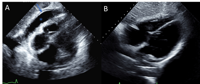
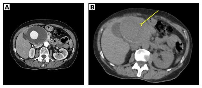
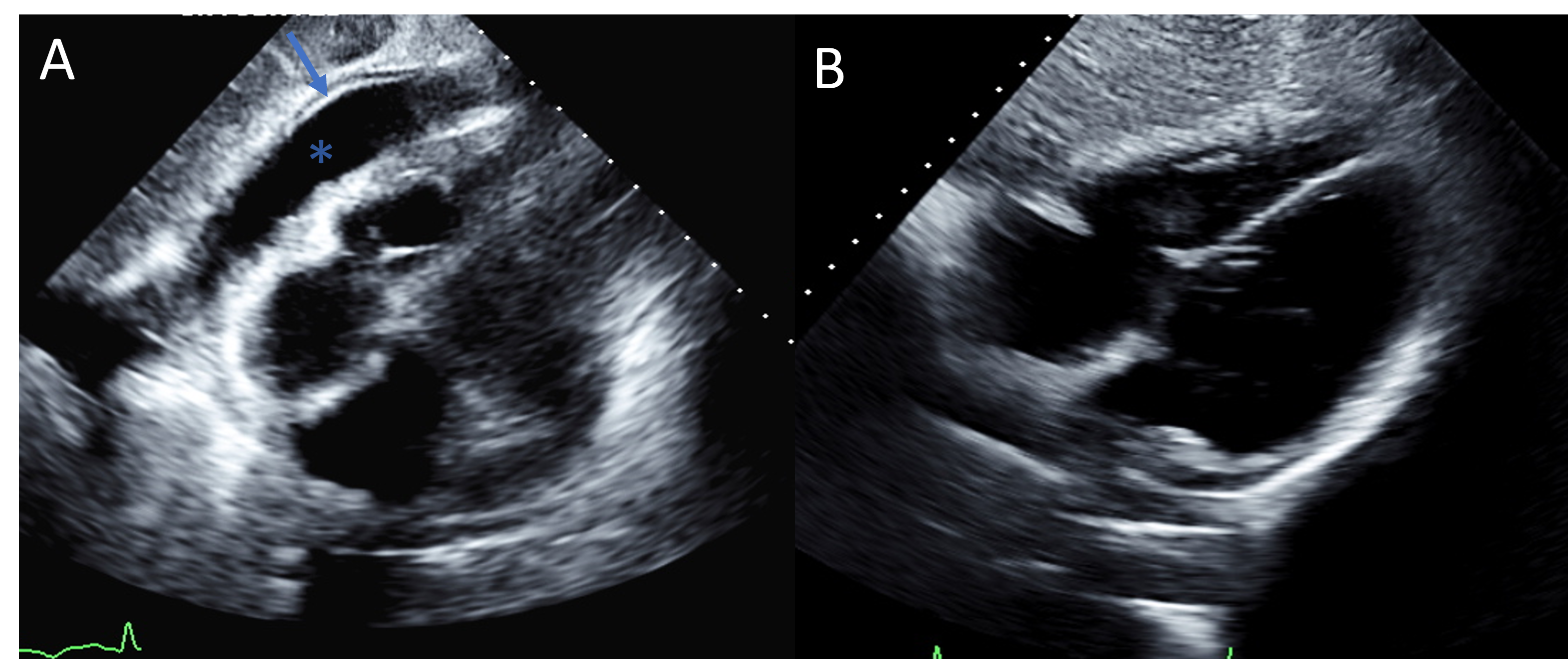
Admission transthoracic echocardiogram in the subcostal view confirmed pericardial thickening and a severe pericardial effusion predominantly localized anterior to the right ventricular free wall (Video 1). A parasternal short-axis view at the level of the great vessels revealed pericardial thickening and a localized pericardial effusion anterior to the right ventricular free wall (Video 2). Echocardiographic changes from admission to the start of medical therapy are shown in Figure 1: pericardial thickening (Figure 1A, arrows) and pericardial effusion (Figure 1A, asterisk) were seen in the subcostal view, and the pericardial effusion was resolved following the initiation of medical therapy (Figure 1B).
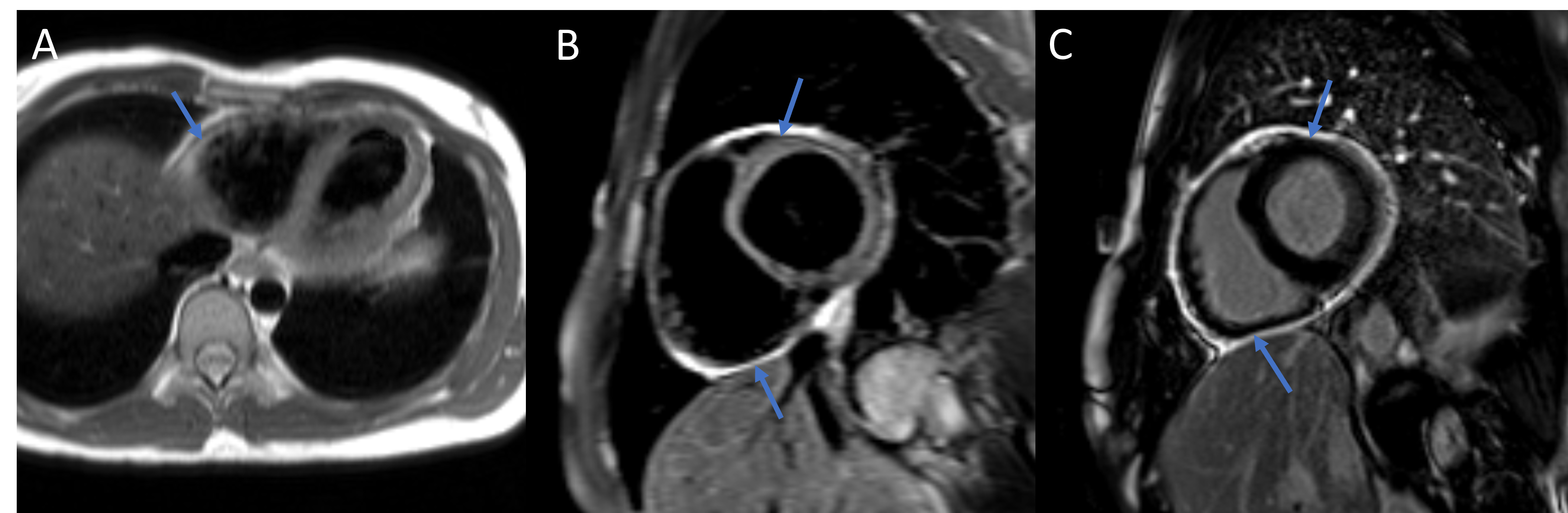
Following clinical improvement with medical therapy, the patient reported recurrent chest pain coinciding with the taper of corticosteroid therapy. An electrocardiogram was performed, showing nonspecific repolarization abnormalities. A cardiac magnetic resonance imaging (CMR) study was planned to complete the evaluation of the chest pain. The CMR showed mild pericardial thickening (up to 4 mm) on black-blood imaging (Figure 2A, arrow). On limited T2-weighted imaging, there was evidence of diffuse pericardial edema (Figure 2B, arrows) and circumferential pericardial enhancement on post contrast imaging (Figure 2C, arrows). On real-time free-breathing imaging, there was no significant evidence of increased ventricular interdependence with respiration (Video 3). These findings were consistent with pericarditis with no definite evidence of constriction. Based on the findings, the steroid dosage was increased and subsequently tapered while a steroid-sparing agent (rilonacept) was initiated, resulting in clear symptomatic improvement. Given the symptomatic improvement, no imaging studies have been performed.
The 2025 European clinical practice guidelines indicate that CMR is recommended in patients with suspected pericarditis when a diagnosis cannot be made using clinical criteria.1 In our case, the CMR allowed for the diagnosis of active pericarditis without myocardial involvement in a patient without definitive clinical signs of pericarditis.
Affiliations and Disclosures
Sirisha Vuppalapati; Nicole Zhang; Angelos Tsekos; Muhammad Ali; Sooyun Caroline Tavolacci, MD; Marcos Ferrandez-Escarabajal, MD; Stamatios Lerakis, MD, PhD
*Ms Vuppalapati and Ms Zhang contributed equally to this work.
Department of Cardiology, Mount Sinai Fuster Heart Hospital, Icahn School of Medicine at Mount Sinai, New York, New York.
Conflicts of interest: The authors report no financial relationships or conflicts of interest regarding the content herein.
Consent statement: The authors confirm that informed consent was obtained from the patient for the publication of the case report.
Address for correspondence: Stamatios Lerakis, MD, PhD, One Gustave L. Levy Place, Box 1030, New York, NY 10029-6574, USA. Email: Stamatios.Lerakis@mountsinai.org
References
- Schulz-Menger J, Collini V, Gröschel J, et al; ESC Scientific Document Group. 2025 ESC Guidelines for the management of myocarditis and pericarditis. Eur Heart J. 2025;46(40):3952-4041. doi:10.1093/eurheartj/ehaf192