Melody Valve Embolization During Transcatheter Pulmonary Valve Replacement
© 2026 HMP Global. All Rights Reserved.
Any views and opinions expressed are those of the author(s) and/or participants and do not necessarily reflect the views, policy, or position of the Journal of Invasive Cardiology or HMP Global, their employees, and affiliates.
J INVASIVE CARDIOL 2026. doi:10.25270/jic/26.00252. Epub August 5, 2026.
A 26-year-old man with a history of tetralogy of Fallot repair and biological pulmonary valve replacement 8 years prior was admitted to our center for a transcatheter pulmonary valve implantation because of severe stenosis of the pulmonary valve conduit. The procedure was performed via the right femoral vein, while the left radial artery was used to exclude left main obstruction.
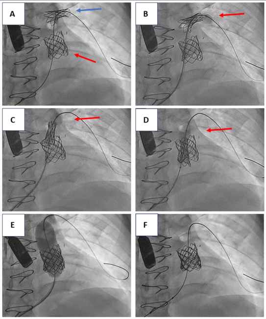
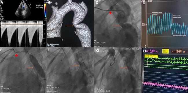
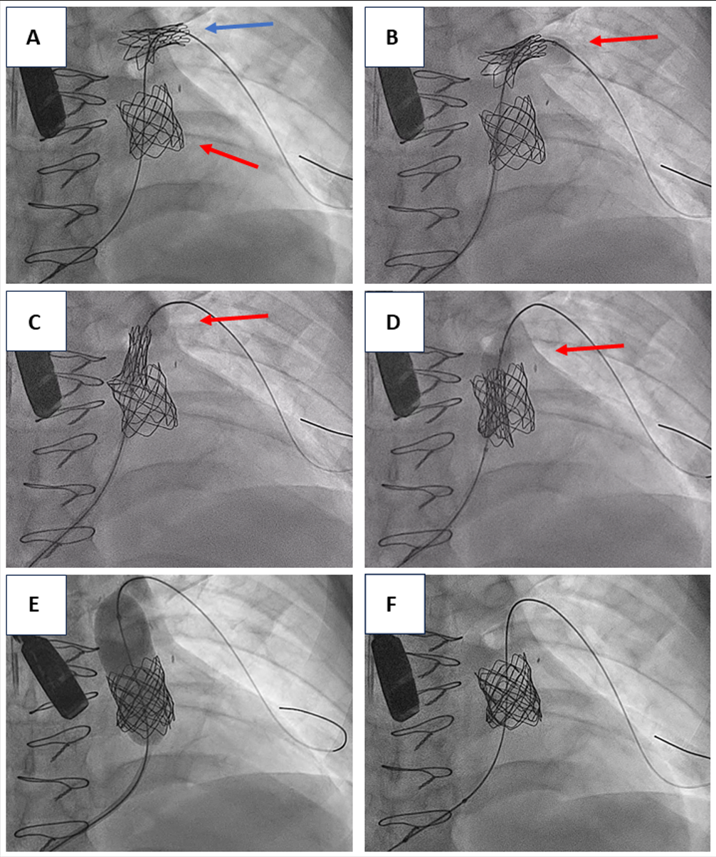
After successful implantation of a Cheatham-Platinum (CP) stent (NuMed) in the pulmonary trunk to act as a landing zone, a 22-mm Melody transcatheter pulmonary valve (Medtronic) mounted in the Ensemble II transcatheter delivery system (Medtronic) was inserted through an 18F sheath in the femoral vein. Advancement of the delivery system into the proper position was challenging, and during repeated attempts the valve mounted into the Balloon-in-Balloon (BIB) delivery system (NuMed) was inadvertently displaced outside the external protective sheath. After pulling the BIB back into the sheath, the valve was partially removed in the upper portion of the delivery balloon. This resulted in embolization of the valve in the left pulmonary artery after the inflation of the outer balloon (Figure A).
In order to retrieve the embolized valve, a 23 x 45-mm high-pressure, noncompliant balloon catheter (Cristal Balloon [BALT Extrusion]) was partially advanced through the Melody valve and was subsequently inflated in low atmospheres (Figure B). The embolized valve was gradually pulled back inside the CP stent (Figures C and D, Video 1). Without deflating the balloon (in order to prevent a second embolization), the Cristal Balloon was fully inflated (Figure E, Video 1) and the Melody valve was successfully expanded inside the pulmonary trunk (Figure F, Video 2). The patient had an uneventful recovery and was discharged the next day with the valve functioning well.
Although Melody valve embolization is a rare complication, interventional cardiologists should be aware of this unexpected complication.
Affiliations and Disclosures
Charalampos Kakderis, MD, MSc; Matthaios Didagelos, MD, PhD; Antonios Kouparanis, MD; Stylianos Daios, MD, PhD; Konstantinos C. Theodoropoulos, MD, PhD; Antonios Ziakas, MD, PhD
From the 1st Cardiology Department, AHEPA University Hospital, Aristotle University of Thessaloniki, Thessaloniki, Greece.
Consent statement: The authors confirm that informed consent was obtained from the patient for intervention described in the manuscript and for the publication thereof, including any and all images.
Disclosures: The authors report no financial relationships or conflicts of interest regarding the content herein.
Address for correspondence: Charalampos Kakderis, MD, 1st Cardiology Department, AHEPA University General Hospital, Kyriakidi 1, Thessaloniki 546 36, Greece. Email: babiskakderis@hotmail.com