STENTYS Xposition S in the Treatment of Chronic Total Artery Occlusion (SXS-CTO Trial)
© 2026 HMP Global. All Rights Reserved.
Any views and opinions expressed are those of the author(s) and/or participants and do not necessarily reflect the views, policy, or position of the Journal of Invasive Cardiology or HMP Global, their employees, and affiliates.
J INVASIVE CARDIOL 2026. doi:10.25270/jic/26.00107. Epub July 23, 2026.
Key Clinical Summary
• Chronic total occlusion PCI may cause late vessel enlargement and stent malapposition; self-expanding drug-eluting stents are designed to better adapt to vessel remodeling than balloon-expandable stents.
• In this randomized pilot trial, self-expanding drug-eluting stents significantly reduced OCT-detected strut malapposition at 6 months vs balloon-expandable stents.
• Despite improved stent apposition, self-expanding stents showed greater neointimal hyperplasia, greater late lumen loss, and numerically higher in-stent restenosis, with no demonstrated clinical benefit.
Abstract
Objectives. Chronic total occlusion (CTO) percutaneous coronary intervention (PCI) can lead to positive vascular remodeling after revascularization, potentially resulting in late stent malapposition. Self-expanding drug-eluting stents (SE-DES) may adapt better to dynamic vessel diameters than balloon-expandable drug-eluting stents (BE-DES), potentially reducing strut malapposition after CTO-PCI. The authors compared stent malapposition at 6 months between SE-DES and BE-DES in CTO-PCI using optical coherence tomography (OCT).
Methods. In this single-center, prospective, randomized controlled trial, 23 patients with CTO were allocated 1:1 to SE-DES (STENTYS Xposition S [STENTYS S.A]) or BE-DES following successful lesion crossing and balloon predilatation. The primary endpoint was the percentage of malapposed stent struts on 6-month OCT. Secondary endpoints included neointimal hyperplasia, late lumen loss, in-stent restenosis (ISR), and clinical outcomes.
Results. At 6 months, OCT showed significantly lower malapposition in the SE-DES group compared with BE-DES (0.0% [0.0-0.0] vs 0.8% [0.0-2.4]; P = .01). SE-DES was associated with greater neointimal thickness (0.3 mm vs 0.1 mm; P < .001) and late lumen loss (0.7 mm vs 0.1 mm; P = .003). ISR occurred more frequently in the SE-DES group (4 vs 1 patients), without reaching statistical significance. No major clinical events were reported in either group.
Conclusions: SE-DES were associated with a statistically significant reduction in strut malapposition compared with BE-DES. However, the absolute malapposition burden was very low in both groups, making the clinical relevance of this imaging finding uncertain. These findings emphasize the importance of individualized stent selection and systematic intracoronary imaging in complex PCI.
Introduction
Chronic total occlusions (CTO) are found in 5% to 10% of patients undergoing coronary angiography. CTO percutaneous coronary intervention (CTO-PCI) is regularly performed with a high success rate, aiming to improve symptoms and, in selected cases, prognosis.1-3
CTO leads to a negative vascular remodeling, resulting in constriction of the underperfused vessel. However, after successful recanalization and restoration of normal coronary flow, positive remodeling occurs with subsequent vessel enlargement, making accurate stent sizing challenging.4
Drug-eluting stents (DES) provide excellent outcomes in terms of in-stent restenosis (ISR) or stent thrombosis. Nevertheless, because of their rigid metallic structure, DES cannot accommodate changes in vessel diameters. Consequently, several studies have shown that late stent malapposition was common after CTO-PCI.5-8
Self-expanding (SE) DES may offer a potential solution to the challenges of positive remodeling and late strut malapposition in CTO recanalization. Indeed, these stents have been used in several clinical studies in different contexts, demonstrating favorable outcomes in terms of stent apposition, ISR, and strut coverage, with an excellent safety profile.9-23
Whether SE-DES improve outcomes compared with balloon-expandable (BE) DES in the setting of CTO-PCI remains uncertain, and whether such morphological findings translate into clinical benefit is unknown. To date, no randomized study has addressed this question. The SXS-CTO trial was therefore designed to compare the incidence of stent malapposition between SE-DES (STENTYS Xposition S [STENTYS S.A]) and BE-DES in patients undergoing CTO-PCI.
Methods
Study design and settings
The SXS-CTO Trial (comparison between BE-DES and SE-DES STENTYS Xposition S in the treatment of chronic total coronary occlusions) was a prospective, parallel group, open-label, randomized controlled single-center trial conducted at Henri Mondor University Hospital, France, between January 2018 and April 2019 that compared SE-DES (STENTYS Xposition S) with BE-DES in the setting of CTO-PCI.
Patients were informed about the study after providing consent to the coronary angiography procedure. Written informed consent was obtained before randomization after detailing all processes of the study prior to inclusion. This study was approved by an ethics committee (CPP – Sud Méditerranée III), and was performed according to the Declaration of Helsinki, Good Clinical Practice guidelines, and French legislation on clinical research. This trial was registered at ClinicalTrials.gov (NCT03563989).
Study population
All patients with a CTO on coronary angiography were screened for eligibility. Inclusion criteria were the presence of a CTO, indication for CTO-PCI confirmed by 2 interventional cardiologists, and target vessel diameter between 2.5 mm (matching the smallest STENTYS stent size) and 4.5 mm (corresponding to the largest BE-DES available in our center).
Exclusion criteria included the presence of significant calcifications, in-stent CTO, known allergy to stent components, or pregnancy/breastfeeding. Patients were also excluded prior to randomization in cases of failed guidewire or balloon crossing of the CTO, or persistent balloon waist during predilation.
Procedure
Patients were admitted 1 day before the intervention. PCI was performed according to standard practice and current guidelines. Two experienced operators worked in tandem. Both antegrade and retrograde approaches were used, except in the absence of collaterals. The choice of approach, guiding catheter, and revascularization strategy was at the operator’s discretion.
After CTO crossing, systematic predilation with noncompliant balloons was performed. Stent selection was guided by angiographic assessment or intracoronary imaging (ICI), as per operator preference. Final optical coherence tomography (OCT) evaluation was strongly recommended. The choice of BE-DES brand was left to the discretion of the operators.
Pharmacological management was standardized in both study arms. Dual antiplatelet therapy (DAPT) consisted of aspirin and a P2Y12 inhibitor (usually clopidogrel). Antiplatelet therapy naïve patients received a loading dose of clopidogrel (600 mg) and/or aspirin (250 mg) the day before PCI. Intravenous unfractionated heparin (100 IU/kg) was administered at the beginning of the procedure, with activated clotting time (ACT) monitored every 30 minutes, targeting a value of greater than 300 seconds.
DAPT was continued for at least 6 months post-procedure. At 6 months, patients underwent follow-up coronary angiography and OCT imaging, in addition to a clinical assessment for adverse events.
Randomization and treatment
Patients were randomized in the catheterization laboratory using a computer-generated sequence following successful CTO crossing and balloon predilation.
The randomization was performed using variable block sizes and was stratified by diabetes to minimize imbalance in this known risk factor for restenosis and adverse clinical outcomes.
Investigational device
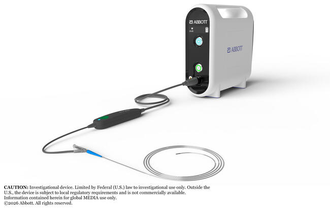
The Xposition S self-apposing stent (STENTYS) is made of a laser-cut nitinol (nickel-titanium alloy) tube. It is compatible with 6F guide catheters and delivered via a rapid exchange delivery system over a standard 0.014-inch guidewire. The device is deployed by withdrawal of a retractable sheath and is available in 4 lengths (17, 22, 27, 37 mm) and 3 diameters: small (2.5-3 mm vessels), medium (3-3.5 mm), and large (3.5-4.5 mm). Due to the mechanical properties of nitinol, the stent can conform to the vessel’s shape and expand beyond nominal diameters: up to 4.0 mm for small, 5.0 mm for medium, and 6.0 mm for large sizes.
Study endpoints and follow-up
The primary endpoint was the percentage of malapposed stent struts at 6 months, assessed by OCT. Secondary endpoints included 6-month adverse clinical events (symptoms including angina [Canadian Cardiovascular Society classification] and dyspnea [New York Heart Association classification], all-cause mortality, and cardiovascular events: cardiovascular death, myocardial infarction, stroke, and target vessel revascularization).
Additional morphological endpoints at 6 months included the following:
- Angiographic parameters: in-stent restenosis (>50% stenosis), minimal luminal diameter (MLD) within the stent and within 5 mm of both stent edges, mean and maximum luminal diameter, late lumen loss (difference in MLD between post-PCI and 6-month angiography), and distal vessel diameter (to assess remodeling);
- OCT parameters: strut coverage, mean/maximum/minimum endoluminal diameter, malapposed surface area, number of patients with malapposed struts of greater than 5%, and dissection area.
Angiographic and OCT assessment
Six-month coronary angiography was performed after intracoronary administration of 1 mg of nitroglycerin.
OCT was systematically performed in the recanalized artery, using a C7 Dragonfly Imaging Catheter (Abbott, formerly St. Jude Medical) with contrast injection and pullback imaging. Data were analyzed using LightLab Imaging software (OCT system software B.0.1 [Abbott, formerly St. Jude Medical]).
All OCT analyses were performed by an independent core laboratory (Krakow Cardiovascular Research Institute [KCRI], Poland) which was blinded to treatment allocation and clinical outcomes.
OCT analysis included the intra-stent zone as well as the proximal and distal edges over 5 mm. Images with poor visibility or distortion were excluded (insufficient blood clearance or the presence of an intraluminal mass that impeded the analysis). Struts located at bifurcations were excluded as they are inherently malapposed. OCT analysis followed the APPOSITION studies protocol, with 1-mm spaced measurements.9 Semi-automated internal lumen contours were therefore generated at 1-mm intervals and corrected manually as needed. Stent apposition was assessed over the entire arterial circumference, as was strut coverage. Malapposition was defined as a strut-wall separation exceeding the manufacturer's reported strut thickness. Parameters collected included arterial and stent morphology, strut apposition, and strut coverage.
OCT during index procedure was recommended. If performed, it was analyzed using the same protocol as follow-up OCT.
Angiographic analysis was performed at baseline and at 6 months by the same independent core laboratory (KCRI, Poland) using the Cardiovascular Angiography Analysis System (CAAS) 5.11.1 software (Pie Medical Imaging).
Statistical analysis
Sample size
Assuming a malapposition rate of 11% (standard deviation [SD]: 10) in the BE-DES group as reported by Heeger et al,24 the analysis of 19 patients per group will allow the detection of a minimal absolute difference of 7% in malapposition rate (malapposition of 4% in the SE-DES group, with an SD of 4) between the 2 arms, with 80% power and a 2-sided alpha risk of 5%. Taking into account a 5% loss to follow-up, randomization of 20 patients per arm will be required. Considering a 20% crossing failure rate, a total of 50 patients will need to be enrolled in the study.
Baseline characteristics were summarized using mean and SD or median and interquartile range (IQR), as appropriate, for quantitative variables and counts and percentages for categorical variables. Analyses were conducted in the intention-to-treat population without any imputation of missing data.
For the primary endpoint and other quantitative endpoints, results were expressed using mean ± SD, or median (IQR), as appropriate, for each arm and compared between arms with Student t-test or Mann-Whitney test, as appropriate. Comparisons between baseline and 6-month follow-up of the quantitative variables were performed using paired Student t-tests or signed-rank tests, as appropriate. For categorical variables, results were expressed using raw numbers and percentages for each arm and compared between arms with chi-square tests or Fisher’s exact tests, as appropriate.
All significance tests were 2-tailed, and the threshold for statistical significance level was set to 5%. No adjustment for multiple comparisons was performed. Therefore, secondary OCT and angiographic endpoints should be interpreted as exploratory. All analyses were performed with Stata software (version 17.0, StataCorp).
Results
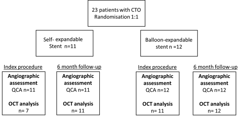
Between August 12, 2017, and September 24, 2019, 23 patients were included and randomized in this trial. Eleven patients were randomized to the SE-DES group and twelve to the BE-DES group. Eighteen patients (78.3%) underwent OCT at the end of the recanalization procedure. At the 6-month follow-up, all 23 patients had both clinical and OCT evaluations (Figure 1). The number of included patients was lower than initially planned because of the manufacturer's market withdrawal of the investigational stent used in this trial.
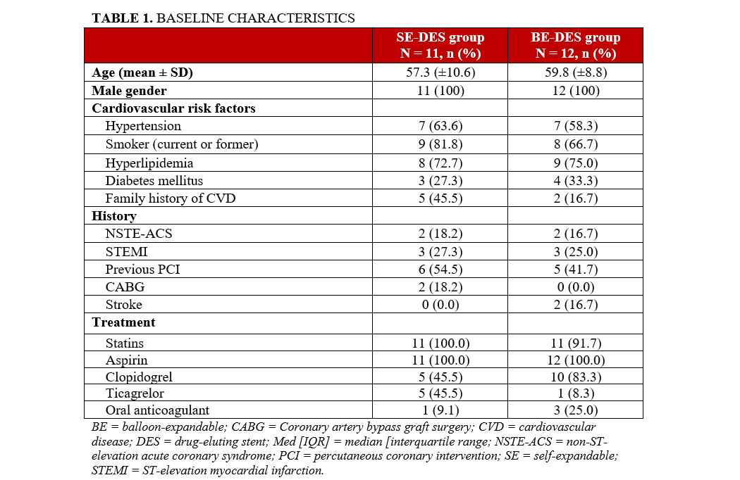
The mean age was 57.3 years in the SE-DES group and 59.8 years in the BE-DES group. Cardiovascular risk factors were comparable between the 2 groups and ventricular function was preserved. Only men were included in both arms (11 in the SE-DES group and 12 in the BE-DES group). (Table 1).
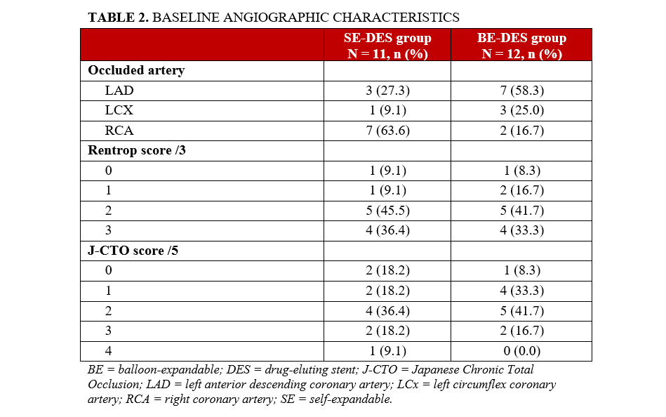
A J-CTO score of 2 or greater was present in 7 patients (63.6%) of the SE-DES group and 7 patients (58.3%) in the BE-DES group. More than half of the lesions were longer than 20 mm (Table 2).
Procedural parameters were comparable between groups, except for procedure time and fluoroscopy time, which were significantly longer in the SE-DES group. A dual injection was used in 20 procedures (87%) typically involving right radial and right femoral access (Table 3).
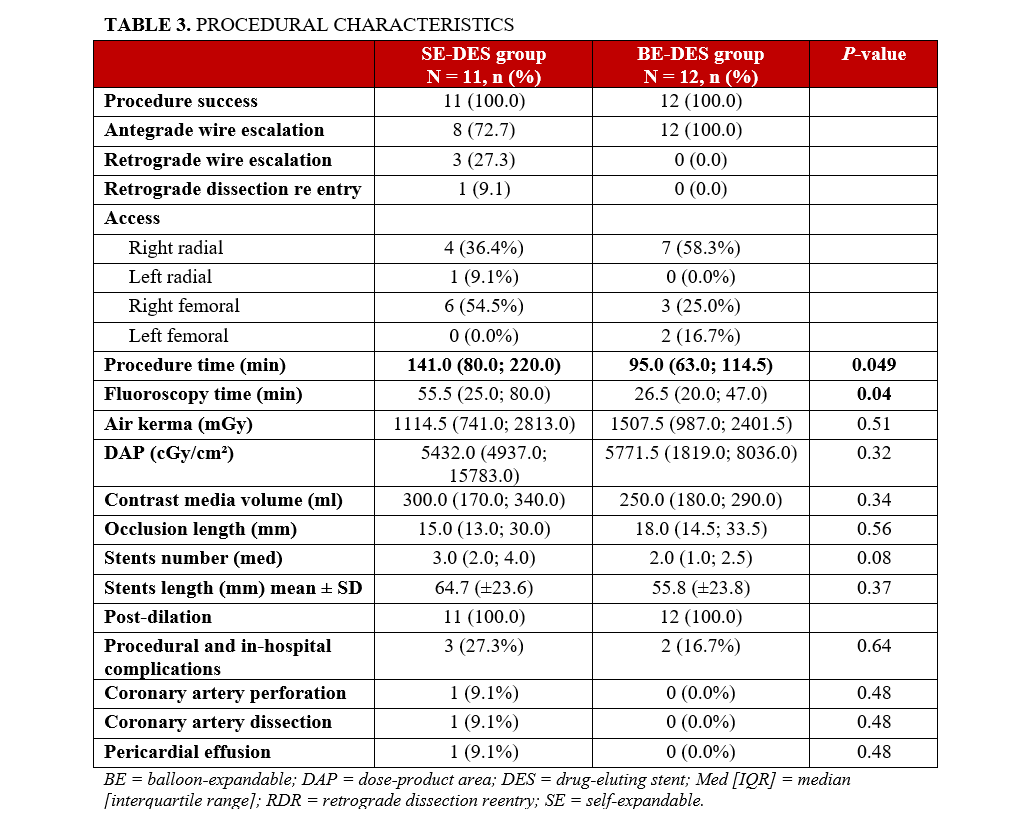
CTO-PCI was mainly performed via the antegrade approach (20 cases, 87.0%). In the SE-DES group, a median of 3.0 [2.0; 4.0] stents were implanted, compared to 2.0 [1.0; 2.5] in the BE-DES group (P = .08). Both pre- and post-dilatation were systematically performed in all 23 cases (Table 3). Detailed stent platform and implantation characteristics, including BE-DES type, drug/polymer platform, strut thickness, diameters, lengths, overlap frequency, and post-dilatation strategy, are reported in the Supplemental Table.
Compared with the BE-DES group, the SE-DES group showed numerically greater procedural complexity, including more frequent right coronary artery (RCA) CTO, greater use of retrograde techniques, longer procedure and fluoroscopy times, and a numerically higher number of implanted stents.
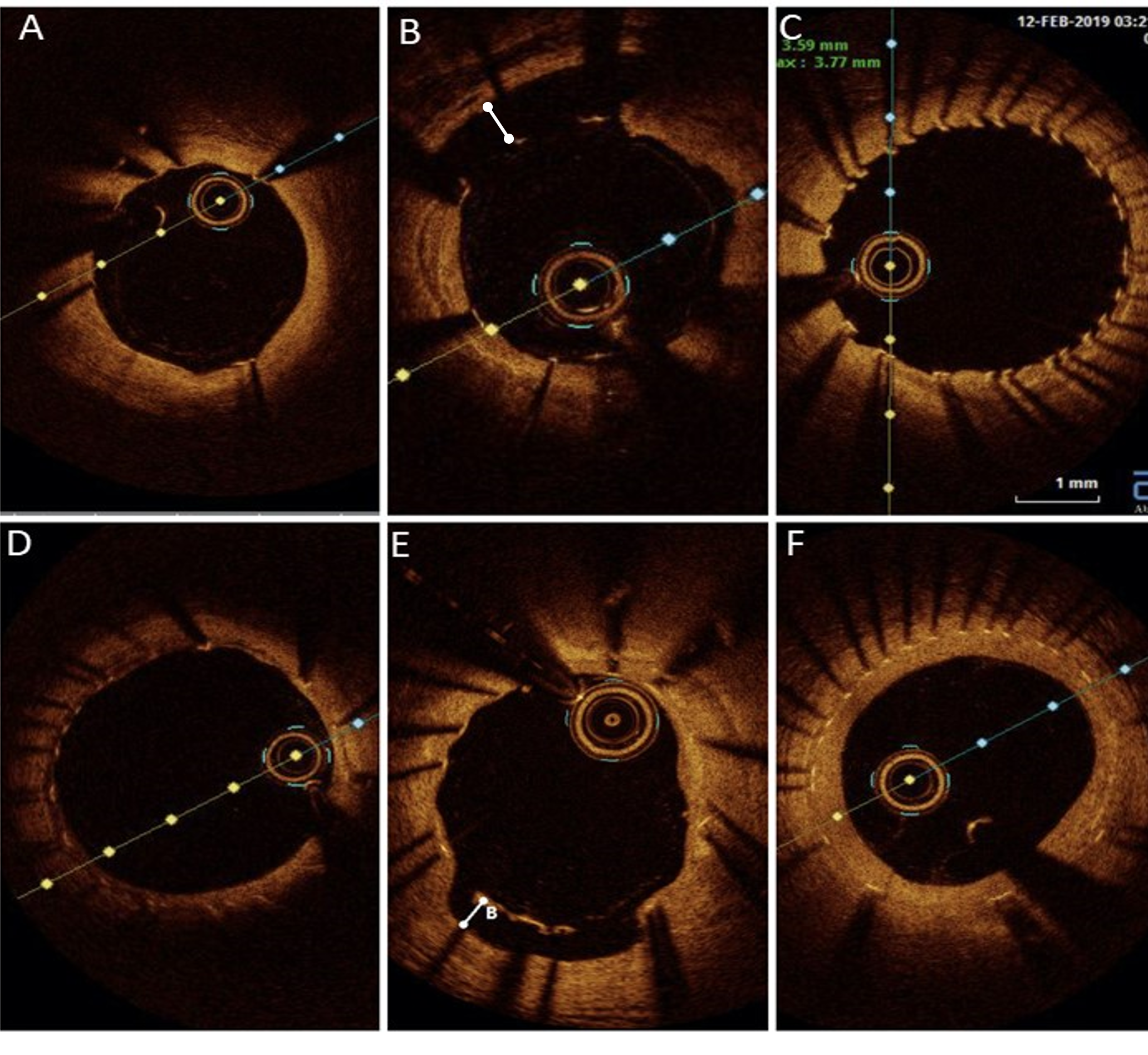
One patient (9.1%) in the SE-DES group experienced a mid-RCA perforation post-dilatation, which was successfully managed with a covered stent. Additionally, 1 dissection (9.1%) and 1 pericardial effusion (9.1%) were reported in the SE-DES group (Figure 2, Table 3).
Six-month clinical, angiographic and OCT results
Six-month follow-up was completed for all patients (N = 23). OCT imaging showed a low percentage of malapposed struts in both groups, with a significantly lower rate in the SE-DES group (0.0% [0.0; 0.0] vs 0.8% [0.0; 2.4], P = .01). All malapposition parameters favored the SE-DES group (Table 4).
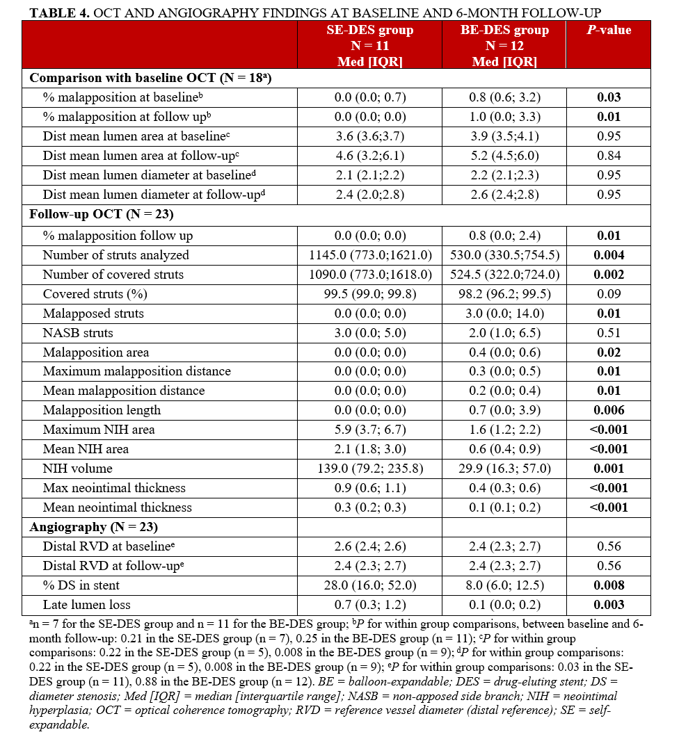
Strut coverage was high in both arms (99.5% [99.0; 99.8] in SE-DES vs 98.2% [96.2; 99.5] in BE-DES, P = .09). SE-DES group showed significantly greater neointimal thickness (0.3 mm [0.2; 0.3] vs 0.1 mm [0.1; 0.2], P < .001), correlating with a greater late lumen loss on angiography (0.7 mm [0.3; 1.2] vs 0.1 mm [0.0; 0.2], P = .003) (Table 4).
Restenosis rates differed between groups, though it did not reach statistical significance (P = .16). In the SE-DES group, 4 patients (36.4%) developed ISR, which was treated with repeat PCI (3 patients, 75.0%) or bypass surgery (1 patient, 25.0%). In the BE-DES group, 1 patient (8.3%) developed ISR, which was managed with a drug-eluting balloon.
Discussion
In this prospective randomized study comparing the STENTYS SE-DES with standard BE-DES for the treatment of CTO, the main findings of this trial can be summarized as follows:
- SE-DES was associated with a statistically significant reduction in OCT-detected stent malapposition at 6 months compared with BE-DES;
- At 6 months, strut malapposition rates remained low in both groups, reflecting effective stent expansion;
- SE-DES was associated with greater neointimal hyperplasia, greater late lumen loss, and a numerically higher rate of ISR.
After CTO recanalization, restored coronary flow induces distal positive remodeling. Since stent sizing is usually based on vessel diameter after balloon predilatation and intracoronary nitrate injection, chronic negative remodeling may lead to stent undersizing at implantation. Subsequent vessel enlargement, particularly at distal edges, may then predispose to late stent malapposition.4 SE-DES were specifically designed to address this challenge, as their ability to adapt continuously to vessel enlargement may reduce the risk of malapposition. SE-DES were evaluated first in the context of ST-elevation myocardial infarction (STEMI) where index vasoconstriction may lead to stent undersizing. In the APPOSITION trials, OCT analyses demonstrated a significantly lower incidence of malapposed struts with SE-DES compared with balloon-expandable stents, while clinical outcomes confirmed a favorable safety profile. SE-DES were later investigated in complex coronary lesions (left main artery, bifurcations, saphenous bypass, coronary aneurysm), showing global favorable procedural outcomes and a low rate of clinical events.6, 14-20,22,23,25 Given the parallels between vessel dynamic changes in STEMI and CTO—both characterized by initial vessel shrinkage followed by positive remodeling—SE-DES can be expected to provide favorable outcomes in the context of CTO-PCI.
In the 2020 study by Vlieger et al, 15 CTOs were recanalized by implanting STENTYS stents. A systematic OCT evaluation was performed at 6 weeks, showing a malapposition rate of 0.9%.21 This rate was even lower in our study (0.1% at 6 months), supporting favorable apposition of the STENTYS stent in CTO-PCI.
In a recent prospective, non-randomized, single-center study, Elborae et al compared 20 patients who underwent CTO PCI using SE-DES with 20 matched control patients who underwent CTO PCI using BE-DES. At 12 months, the SE-DES group had fewer malapposed struts (0% [IQR 0%-0%] vs 4.5% [IQR 0%-20%]; P = .001) and uncovered struts (0.08% [IQR 0%-1.6%] vs 8.2% [IQR 0%-16%]; P = .001). However, the SE-DES group had a higher rate of major adverse cardiovascular events, driven by clinically indicated target lesion revascularization (45% vs 15%; P = .038). A major limitation of this study is that, even in the SE-DES group, many patients also received BE-DES implantation, especially in distal segments where positive remodeling most commonly occurs.26
It is noteworthy that, in both groups in our study, the absolute number and percentage of malapposed struts were very low, even immediately after the procedure. Moreover, malapposition rates in our study were substantially lower than those previously reported in the literature. 21, 24, 26-28
In a study by Sherbet et al, OCT was performed in 62 patients at 8 months after CTO-PCI; results showed that 9% of stent struts were malapposed and 2.8% of struts were uncovered by endothelium. Malapposition was observed in 50 out of the 62 patients.27 In the study by Heeger et al, 19 patients with 20 CTO lesions underwent follow-up OCT at 6.5 ± 2.1 months. At follow-up, 20.2% ± 16.2% of struts per patient were uncovered but apposed, whereas 10.9% ± 10.3% were uncovered and malapposed.24
In the CONSISTENT-CTO study, the OCT percentage of malapposition was 2.5% at 12 months.28 These results are consistent with our observations and likely reflect current practices. Indeed, ICI has been shown to improve stent morphological parameters in complex procedures. In our study, 78% of patients underwent OCT imaging at the end of the recanalization procedure, allowing for stent optimization. Similarly, intracoronary ultrasound was used in 90% of patients in CONSISTENT-CTO and 75% of patients in the study by Elborae et al.26,28
In contemporary registries, ICI during CTO-PCI does not exceed 38%, with IVUS preferred over OCT despite OCT’s higher sensitivity for detecting malapposition.28 In the group of 18 patients who underwent OCT during the index procedure, there was no significant difference in terms of malapposition between the index procedure and the follow-up at 6 months; this demonstrates that there were no late malappositions, and also suggests that the implanted stents were appropriately sized and that they benefited from post-dilatations using adequately-sized balloons. These findings support the broader use of ICI for stent optimization in CTO-PCI.
Although the difference in malapposed struts reached statistical significance, the clinical relevance of this morphological difference remains uncertain. OCT studies of stent thrombosis have identified stent malapposition as one of the potential mechanisms associated with late and very late stent thrombosis.29 However, thrombotic risk appears to depend less on the mere presence of isolated malapposed struts than on the extent of malapposition and its association with other adverse OCT features, such as uncovered struts or neoatherosclerosis. In the present study, the absolute burden of malapposition was extremely low in both groups, with a median percentage of malapposed struts of 0.0% in the SE-DES group vs 0.8% in the BE-DES group. Accordingly, despite its statistical significance, this OCT-detected morphological finding should not be interpreted as evidence of the clinical superiority of SE-DES. Moreover, the study was neither designed nor powered to assess clinical benefit. This cautious interpretation is further supported by the greater neointimal hyperplasia and late lumen loss observed in the SE-DES group, together with a numerical increase in ISR.
OCT analysis showed greater strut coverage with SE-DES, but this was offset by greater neointimal thickness and increased late lumen loss at 6 months. These findings, consistent with APPOSITION I,9,25 suggest that the morphological benefit in apposition may be counterbalanced by excess neointimal proliferation with a numerical, though not statistically significant, increase in ISR in the SE-DES group.9,26
The greater neointimal hyperplasia, late lumen loss, and numerical increase in ISR observed in the SE-DES group should be interpreted cautiously, as anatomical and procedural imbalances between the groups may have contributed to these findings. Patients in the SE-DES group more frequently had RCA CTOs, more often underwent retrograde procedures, and received a numerically greater number of stents. Longer-stented segments, stent overlap, and greater procedural complexity may themselves promote neointimal proliferation and restenosis. Given the very small sample size, formal multivariable adjustment would not have been statistically reliable; therefore, no causal relationship between the stent platform and these findings can be established.
Nevertheless, a device-related mechanism remains plausible. The STENTYS stent is made of nitinol, crimped onto a balloon, and covered with a sheath. Nitinol is a shape-memory alloy with significant radial force. If the deployed stent is constrained by plaque and cannot reach its normal diameter, it will exert continuous force on the artery over the long term. This persistent mechanical stress, combined with the acute injury induced by balloon inflation and sheath peeling during deployment, may contribute to the proliferative intimal hyperplasia frequently observed during follow-up of STENTYS stents.
Beyond OCT-based stent assessment, coronary imaging plays an important role throughout CTO-PCI. Coronary computed tomography angiography can provide a noninvasive 3-dimensional assessment of CTO anatomy, including lesion length, vessel course, proximal and distal cap morphology, calcification, tortuosity, and distal vessel quality. These features may help anticipate procedural complexity, guide strategy selection, and identify lesions at higher risk of failure or complications.30 Intravascular imaging remains complementary: IVUS may assist with cap identification, wire position confirmation, ambiguity resolution, and stent sizing, whereas OCT provides high-resolution assessment of strut apposition, tissue coverage, edge dissection, and neointimal response.31,32 In this context, the very low malapposition rates observed in both arms may partly reflect frequent intravascular imaging use and systematic post-dilatation.
Limitations
To the best of our knowledge, this is the first randomized study to evaluate SE-DES in the setting of CTO-PCI. Several limitations should be acknowledged. First, this was a single-center, exploratory, pilot mechanistic study focused on OCT-derived morphological endpoints. Only 23 of the 50 planned patients were randomized because recruitment had to be terminated prematurely following the cessation of STENTYS’ commercial activities. This small sample size markedly limits statistical power, precludes any reliable assessment of clinical outcomes, and requires cautious interpretation of secondary endpoints. Second, randomization was performed only after successful CTO crossing and balloon predilatation; therefore, the findings apply only to successfully crossed and adequately predilated CTO lesions and cannot be generalized to an all-comer CTO-PCI population. Third, multiple OCT and angiographic endpoints were analyzed without adjustment for multiplicity, and secondary findings should therefore be considered exploratory. Fourth, the higher neointimal hyperplasia and restenosis signal observed in the SE-DES group may have been influenced by anatomical and procedural imbalances, including RCA predominance, greater use of retrograde techniques, longer procedure and fluoroscopy times, and a numerically higher number of implanted stents. Given the small sample size, multivariable adjustment was not statistically appropriate. Fifth, the BE-DES comparator reflected operator discretion and included different stent platforms, introducing heterogeneity in drug, polymer, strut thickness, radial strength, and mechanical properties. Finally, only men were included, which limits the generalizability of the findings to women.
Conclusions
In the SXS-CTO randomized trial, SE-DES were associated with a statistically significant reduction in OCT-detected strut malapposition compared with BE-DES. However, malapposition was rare in both groups, and the absolute difference was small, making the clinical relevance of this morphological difference uncertain. Moreover, this modest apposition advantage was accompanied by greater neointimal hyperplasia, greater late lumen loss, and a numerical increase in ISR in the SE-DES group. These findings suggest that, in CTO-PCI performed with frequent intravascular imaging guidance and systematic post-dilatation, SE-DES may not provide a clinically meaningful advantage over contemporary BE-DES. This study further supports the role of ICI in optimizing stent implantation in complex coronary interventions.
Affiliations and Disclosures
Madjid Boukantar, MD1; Oueies Labidi, MD1,2; Patrick Zamora, MD1; Romain Gallet, MD, PhD1; Tony Truong, MD1; Nicolas Lellouche, MD, PhD1; Nadia Oubaya, MD3,4; Pierre Andre Natella, MD3,4; Philippe Le Corvoisier, MD5; Paul Mathieu Chiaroni, MD1; Emmanuel Teiger, MD, PhD1
From the 1Interventional Cardiology Department, Henri Mondor Hospital, AP-HP, Créteil, France; 2Cardiology Department, Bicêtre Hospital, AP-HP, Le Kremlin-Bicêtre, France; 3Université Paris-Est Créteil, INSERM, IMRB, Créteil, France; 4AP-HP, Hôpitaux Henri-Mondor, Department of Public Health, Créteil, France; 5INSERM, Centre d'Investigation Clinique 1430, AP-HP, Hôpitaux Universitaires Henri Mondor, Université Paris-Est Créteil, Créteil, France.
Disclosures: The authors report no financial relationships or conflicts of interest regarding the content herein.
Funding: The study was sponsored by Assistance Publique–Hôpitaux de Paris (Délégation à la Recherche Clinique et à l'Innovation).
Consent statement: The authors confirm that informed consent was obtained from the patient(s) for the study and interventions described in the manuscript and to the publication of their data, including any and all images.
Data availability statement: The data supporting the findings of this study are available upon reasonable request.
Address for correspondence: Madjid Boukantar, MD, Interventional Cardiology Department, Henri Mondor Hospital, AP-HP, 1 Rue Gustave Eiffel, Créteil 94000, France. Email: madjid.boukantar@aphp.fr
References
1. Boukantar M, Loyeau A, Gallet R, et al. Angiography and percutaneous coronary intervention for chronic total coronary occlusion in daily practice (from a large French registry [CARDIO-ARSIF]). Am J Cardiol. 2019;124(5):688-695. doi:10.1016/j.amjcard.2019.05.062
2. Hoebers LP, Claessen BE, Dangas GD, Råmunddal T, Mehran R, Henriques JP. Contemporary overview and clinical perspectives of chronic total occlusions. Nat Rev Cardiol. 2014;11(8):458-469. doi:10.1038/nrcardio.2014.74
3. Marechal P, Gach O, Davin L, et al. Why to treat a total chronic coronary occlusion? Article in French. Rev Med Suisse. 2017;13(571):1406-1409.
4. Park JJ, Chae IH, Cho YS, et al. The recanalization of chronic total occlusion leads to lumen area increase in distal reference segments in selected patients: an intravascular ultrasound study. JACC Cardiovasc Interv. 2012;5(8):827-836. doi:10.1016/j.jcin.2012.05.004
5. Verheye S, Grube E, Ramcharitar S, et al. First-in-man (FIM) study of the Stentys bifurcation stent--30 days results. EuroIntervention. 2009;4(5):566-571. doi:10.4244/eijv4i5a96
6. Naber CK, Pyxaras SA, Nef H, et al. Final results of a self-apposing paclitaxel-eluting stent fOr the PErcutaNeous treatment of de novo lesions in native bifurcated coronary arteries study. EuroIntervention. 2016;12(3):356-358. doi:10.4244/EIJY15M06_02
7. Lu H, Bekker RJ, Grundeken MJ, et al. Five-year clinical follow-up of the STENTYS self-apposing stent in complex coronary anatomy: a single-centre experience with report of specific angiographic indications. Neth Heart J. 2018;26(5):263-271. doi:10.1007/s12471-018-1111-7
8. Smolka G, Wańha W, Roleder T, Pluta A, Ochała A. Treatment of left main coronary artery stenosis with the STENTYS self-expandable drug-eluting stent - a pilot registry. Postepy Kardiol Interwencyjnej. 2014;10(4):226-230. doi:10.5114/pwki.2014.46941
9. Amoroso G, van Geuns RJ, Spaulding C, et al. Assessment of the safety and performance of the STENTYS self-expanding coronary stent in acute myocardial infarction: results from the APPOSITION I study. EuroIntervention. 2011;7(4):428-436. doi:10.4244/EIJV7I4A71
10. Nakatani S, Onuma Y, Ishibashi Y, et al. Incidence and potential mechanism of resolved, persistent and newly acquired malapposition three days after implantation of self-expanding or balloon-expandable stents in a STEMI population: insights from optical coherence tomography in the APPOSITION II study. EuroIntervention. 2015;11(8):885-894. doi:10.4244/EIJY15M11_01
11. Koch KT, Grundeken MJ, Vos NS, et al. One-year clinical outcomes of the STENTYS Self-Apposing¨ coronary stent in patients presenting with ST-segment elevation myocardial infarction: results from the APPOSITION III registry. EuroIntervention. 2015;11(3):264-271. doi:10.4244/EIJY15M02_08
12. Lu H, Grundeken MJ, Vos NS, et al. Clinical outcomes with the STENTYS self-apposing coronary stent in patients presenting with ST-segment elevation myocardial infarction: two-year insights from the APPOSITION III (A Post-Market registry to assess the STENTYS self-exPanding COronary Stent In AcuTe MyocardIal InfarctiON) registry. EuroIntervention. 2017;13(5):e572-e577. doi:10.4244/EIJ-D-16-00676
13. van Geuns RJ, Yetgin T, La Manna A, et al. STENTYS Self-Apposing sirolimus-eluting stent in ST-segment elevation myocardial infarction: results from the randomised APPOSITION IV trial. EuroIntervention. 2016;11(11):e1267-e1274. doi:10.4244/EIJV11I11A248
14. Vlieger S, Simsek C, Balland A, et al. Dutch multicenter experience using the STENTYS Xposition S self-apposing stent in complex coronary lesions. Catheter Cardiovasc Interv. 2019;94(7):966-971. doi:10.1002/ccd.28192
15. IJsselmuiden AJJ, Simsek C, van Driel AG, et al. Comparison between the STENTYS self-apposing bare metal and paclitaxel-eluting coronary stents for the treatment of saphenous vein grafts (ADEPT trial). Neth Heart J. 2018;26(2):94-101. doi:10.1007/s12471-017-1066-0
16. Briguori C, Tamburino C, Jessurun GAJ, et al. Prospective evaluation of drug eluting self-apposing stent for the treatment of unprotected left main coronary artery disease: 1-year results of the TRUNC study. Catheter Cardiovasc Interv. 2020;96(2):E142-E148. doi:10.1002/ccd.28584
17. Briguori C, Visconti G, Donahue M, et al. The STENTYS® paclitaxel-eluting stent in the treatment of unprotected distal left main. Catheter Cardiovasc Interv. 2015;86(3):E131-E139. doi:10.1002/ccd.25874
18. Cortese B, Montefusco A, D'Ascenzo F, et al. Clinical performance of a dedicated self-apposing stent for the treatment of left main stem disease. Results of the left Main AngioplasTy wIth a Self-apposing StEnt - the MATISSE study. Cardiovasc Revasc Med. 2018;19(7 Pt B):831-836. doi:10.1016/j.carrev.2018.03.005
19. Kumar N, Kandan SR, Rahbi H, et al. Single-centre experience of STENTYS Xposition S in treatment of left main stem lesions. Expert Rev Med Devices. 2017;14(7):577-582. doi:10.1080/17434440.2017.1344094
20. Siry M, Sukiennik A, Weilenmann D, et al. TCT-470 Worldwide everyday practice registry assessing the Xposition S self-apposing stent in challenging lesions with vessel diameter variance (SIZING registry). J Am Coll Cardiol. sept 2018;72(13_Supplement):B189. doi:10.1016/j.jacc.2018.08.1642
21. Vlieger S, Simsek C, de Jong-Salentijn EM, et al. Increase in coronary lumen area and stent apposition after treatment of CTO using a coronary self-expanding stent. JACC Cardiovasc Interv. 2020;13(4):537-539. doi:10.1016/j.jcin.2019.09.031
22. Mielczarek MJ, Michalski T, Wańha W, et al. Long-term safety and efficacy of self-apposing Stentys drug-eluting stent in left main stem percutaneous coronary intervention: final results of multicentre LM-STENTYS registry. Cardiol J. 2025;32(3):239-247. doi:10.5603/cj.101925
23. Siry M, Duymaz B, Biesenberger S, Siry D, Kammerer V, May AE. Self-expanding versus balloon expanding coronary stents in intervention of the degenerated saphenous vein graft: Memmingen coronary artery bypass stenosis trial (MECAST). J Interv Cardiol. 2023;2023:9412132. doi:10.1155/2023/9412132
24. Heeger CH, Busjahn A, Hildebrand L, et al. Delayed coverage of drug-eluting stents after interventional revascularisation of chronic total occlusions assessed by optical coherence tomography: the ALSTER-OCT-CTO registry. EuroIntervention. 2016;11(9):1004-1012. doi:10.4244/EIJY14M10_01
25. Lu H, De Winter RJ, Koch KT. The STENTYS self-apposing stent technology in coronary artery disease: literature review and future directions. Expert Rev Med Devices. 2018;15(7):479-487. doi:10.1080/17434440.2018.1491305
26. Elborae A, Hassan M, Meguid MA, et al. Self-apposing stents in coronary chronic total occlusions: a pilot study. Heart Lung Circ. 2024;33(4):500-509. doi:10.1016/j.hlc.2024.01.014
27. Sherbet DP, Christopoulos G, Karatasakis A, et al. Optical coherence tomography findings after chronic total occlusion interventions: insights from the "AngiographiC evaluation of the everolimus-eluting stent in chronic Total occlusions" (ACE-CTO) study (NCT01012869). Cardiovasc Revasc Med. 2016;17(7):444-449. doi:10.1016/j.carrev.2016.04.002
28. Walsh SJ, Hanratty CG, McEntegart M, et al. Intravascular healing is not affected by approaches in contemporary CTO PCI: the CONSISTENT CTO study. JACC Cardiovasc Interv. 2020;13(12):1448-1457. doi:10.1016/j.jcin.2020.03.032
29. Souteyrand G, Amabile N, Mangin L, et al; PESTO Investigators. Mechanisms of stent thrombosis analysed by optical coherence tomography: insights from the national PESTO French registry. Eur Heart J. 2016;37(15):1208-1216. doi:10.1093/eurheartj/ehv711
30. Panuccio G, Werner GS, De Rosa S, et al. Impact of coronary calcium patterns on procedural outcomes in CTO-PCI: a computed tomography-based multicenter study. Cardiovasc Interv Ther. 2026;41(1):76-88. doi:10.1007/s12928-025-01200-y
31. Panuccio G, Abdelwahed YS, Carabetta N, Landmesser U, De Rosa S, Torella D. The role of coronary imaging in chronic total occlusions: applications and future possibilities. J Cardiovasc Dev Dis. 2024;11(9):295. doi:10.3390/jcdd11090295
32. Brilakis ES, Mashayekhi K, Tsuchikane E, et al. Guiding principles for chronic total occlusion percutaneous coronary intervention. Circulation. 2019;140(5):420-433. doi:10.1161/CIRCULATIONAHA.119.039797