3D-Printed Implant for Charcot Ankle Reconstruction: A Limb Salvage Case Report
A staged limb salvage approach using a 3D-printed implant achieved stable alignment and progressive fusion in a patient with severe Charcot ankle neuroarthropathy and significant bone loss. Despite postoperative hardware loosening requiring removal, the patient remained pain-free, independently ambulatory, and returned to full-time work at 1-year follow-up.
Key Takeaways
- 3D-printed implants may offer a reconstructive option for severe Charcot ankle deformity with substantial bone loss. In this case, the surgeons incorporated the implant into a staged reconstruction to address the osseous defect, restore alignment, and support tibiotalocalcaneal arthrodesis.
- Hardware complications do not necessarily preclude successful limb salvage. Approximately 4 months after reconstruction, the patient developed hardware irritation and loosening requiring removal; however, the infectious workup was negative, and the joint maintained alignment and progressive fusion.
- Functional outcomes at 1 year were favorable. The patient had a stable, plantigrade foot without recurrent ulceration or skin breakdown, reported no pain with ambulation, walked independently in a CROW boot, and returned to full-time work.
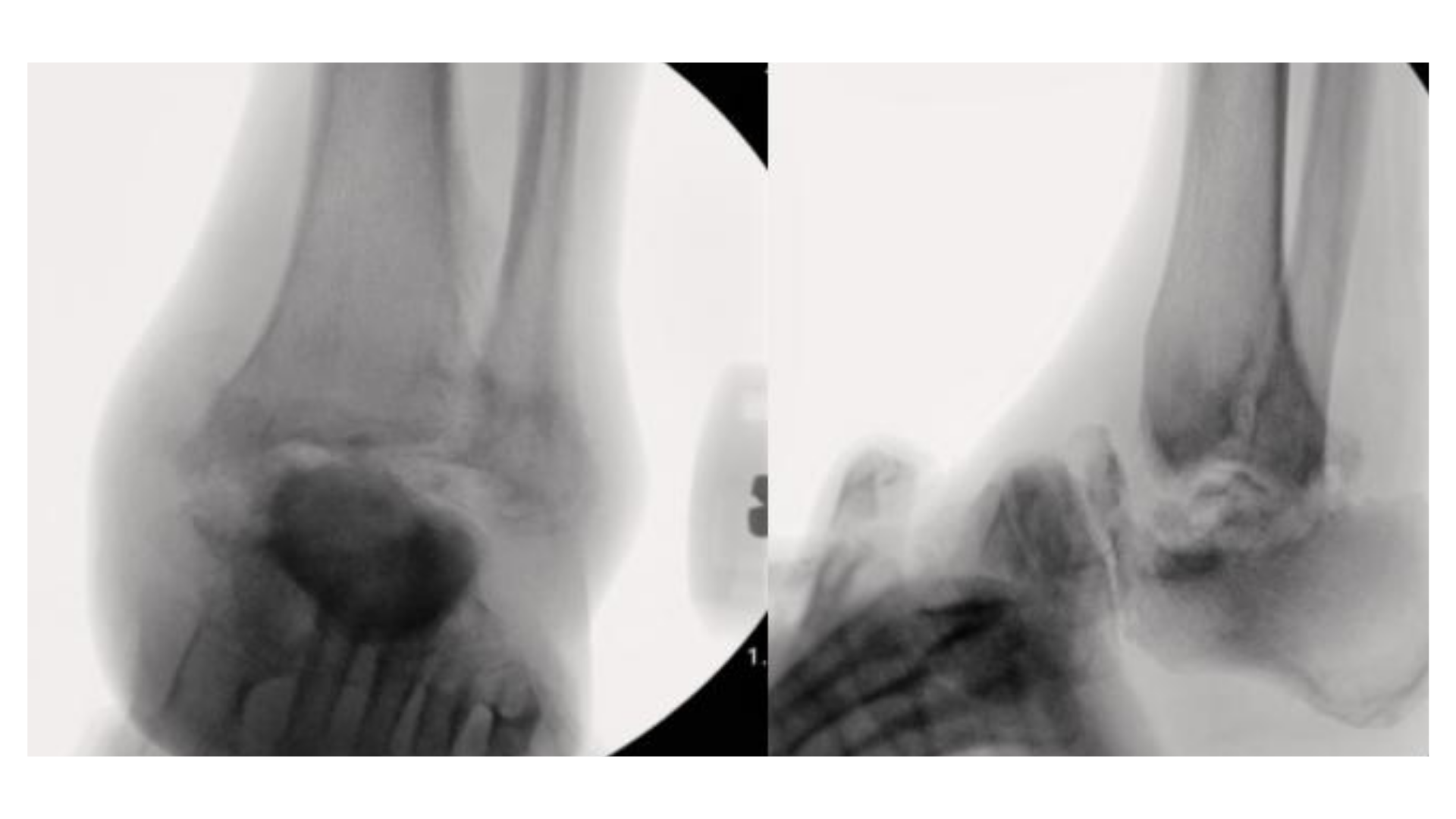
Charcot neuroarthropathy is a peripheral and autonomic neuropathic condition that typically presents initially as a hyperemic event.1 The diagnosis is often clinical, stemming from patient symptoms demonstrating lack of sensation, possible presence of ulceration, and presence of foot deformity. The earliest clinical manifestation of Charcot neuropathy is usually persistent swelling with discomfort, often the main reason the patient seeks medical treatment. It can also present as an acute or chronic condition, with clinical features depending on the nature of the presentation.2 Although the midfoot is more commonly affected by Charcot, cases involving the ankle demonstrate far inferior to results compared to those involving the midfoot.3
Optimal management of Charcot ankle deformity remains controversial for both conservative and surgical treatment. There is no consensus for management of ankle Charcot neuropathy, nor guidelines to define the proper stage for surgical intervention. Offloading orthoses, such as total contact casts, braces, or Charcot restraint orthotic walkers (CROWs), are widely thought to be the most effective initial treatment methods.4 Surgical management can include external fixation, tibiotalocalcaneal fusion, or a combination of both.
In a study of 117 patients, 70 patients underwent intramedullary fixation while 47 underwent external fixation for a tibiotalocalcaneal arthrodesis. The study concluded that intramedullary nails could yield better results than external fixation for the procedure.5 Potential complications for both procedures include infection, delayed union, and nonunion. The researchers found that use of an intramedullary nail yielded double the fusion rate compared to external fixation, with an average of 5 weeks less time to heal.5 An additional complication noted in a separate study comparing TTC, ankle, pantalar, and STJ fusions was implant failure, which demonstrated a rate of 30% on a total of 44 patients.6
Recent advances in 3D printing technology have allowed surgeons to use new approaches in conjunction with traditional fixation, to improve long term outcomes. The Arthrocube (Stryker) implant is composed of a specialized titanium alloy and can potentially help surgeons address large osseous defects and help maintain limb length. In our experience, the implant provides structural stability to assist in improving alignment, while also serving as a scaffold for bone ingrowth, and potentially decreasing risk of nonunion. The theoretical advantage of 3D printing is the seemingly limitless customizability in size, shape, and fixation options.7
This report demonstrates a staged limb salvage approach utilizing circular external fixation followed by utilizing such a 3D implant for reconstruction of a progressive ankle Charcot neuroarthropathy. In addition, the case highlights postoperative hardware complications with eventual successful fusion and function at 1-year follow-up.
Case Report
A 42-year-old male with a past medical history significant for type 2 diabetes mellitus (A1c 12.9), peripheral neuropathy, hyperlipidemia, and body mass index (BMI) of 36, presented with progressive left ankle degeneration and instability. He was initially referred by a physician from an outside facility, after failing conservative treatment modalities, displaying progressive breakdown of the rearfoot despite immobilization with a CROW walker. The patient presented to our office with active Charcot clinical symptoms including erythema, edema, and increased temperature gradient compared to the contralateral extremity. Pedal pulses were palpable to the left lower extremity with adequate capillary refill. The patient had significantly diminished protective sensation to the left foot. There was significant instability of the left ankle, with great anterior translation via anterior drawer test and frontal plane instability on the talar tilt test. Sagittal plane motion of the left ankle was limited, likely secondary to impingement of calcaneus on the tibia. Limb length discrepancy was also noted, secondary to degeneration of the talus. No open wounds were noted to the left lower extremity.
Due to the nature of the deformity, with progression of osseous destruction and instability of the ankle, we advised and planned for a staged surgical approach to attempt limb salvage. We advised the patient to meet with his primary care doctor and endocrinologist for glycemic control before the surgery. There was a 3-month time frame from the initial visit to the first stage of the surgery, in which the patient underwent medical optimization. During this period, the team instructed the patient to remain strictly non-weight-bearing with immobilization to the left lower extremity. The patient did adhere to the preoperative protocol.
Notes on the Surgical Technique
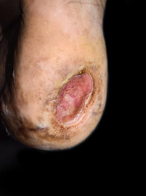
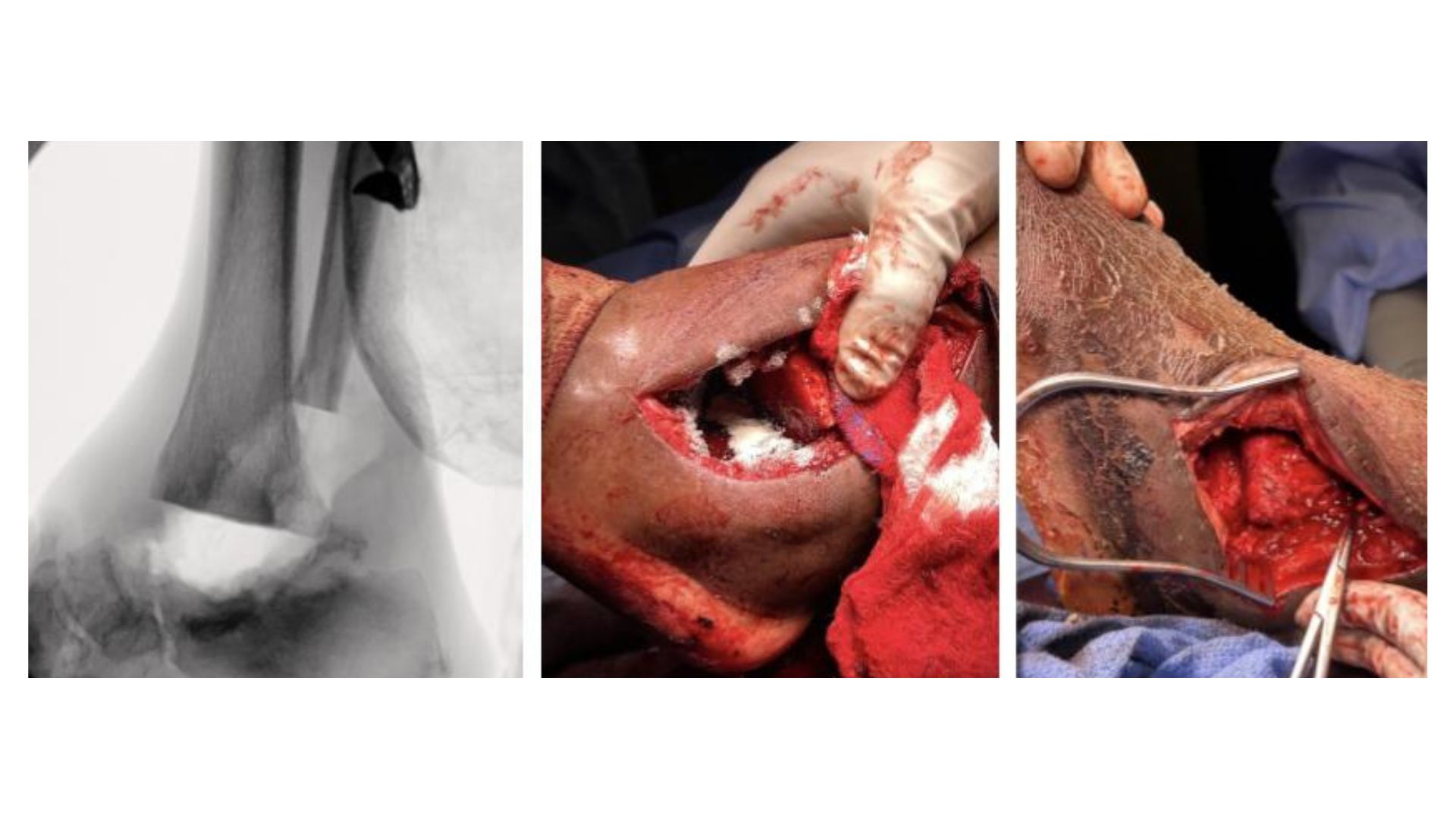
Stage 1: External fixation, bone biopsy, and joint resection (May 2025). The patient underwent resection of nonviable bone, including the distal fibula and fragments of the talus. Resection and removal of the distal fibula allowed access to the ankle joint. We visually noted complete degeneration of the talus, with small fragments remaining. The distal tibia also showed degenerative changes, while the articular surfaces of the calcaneus appeared healthy. We prepared the distal tibia and calcaneus for interposition of the resected fibula (Figures 1 and 2).
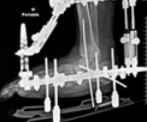
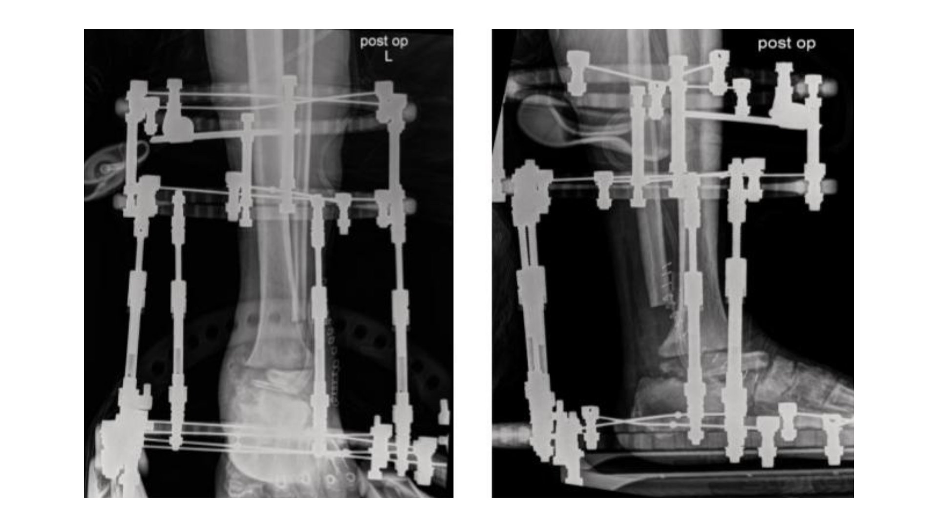
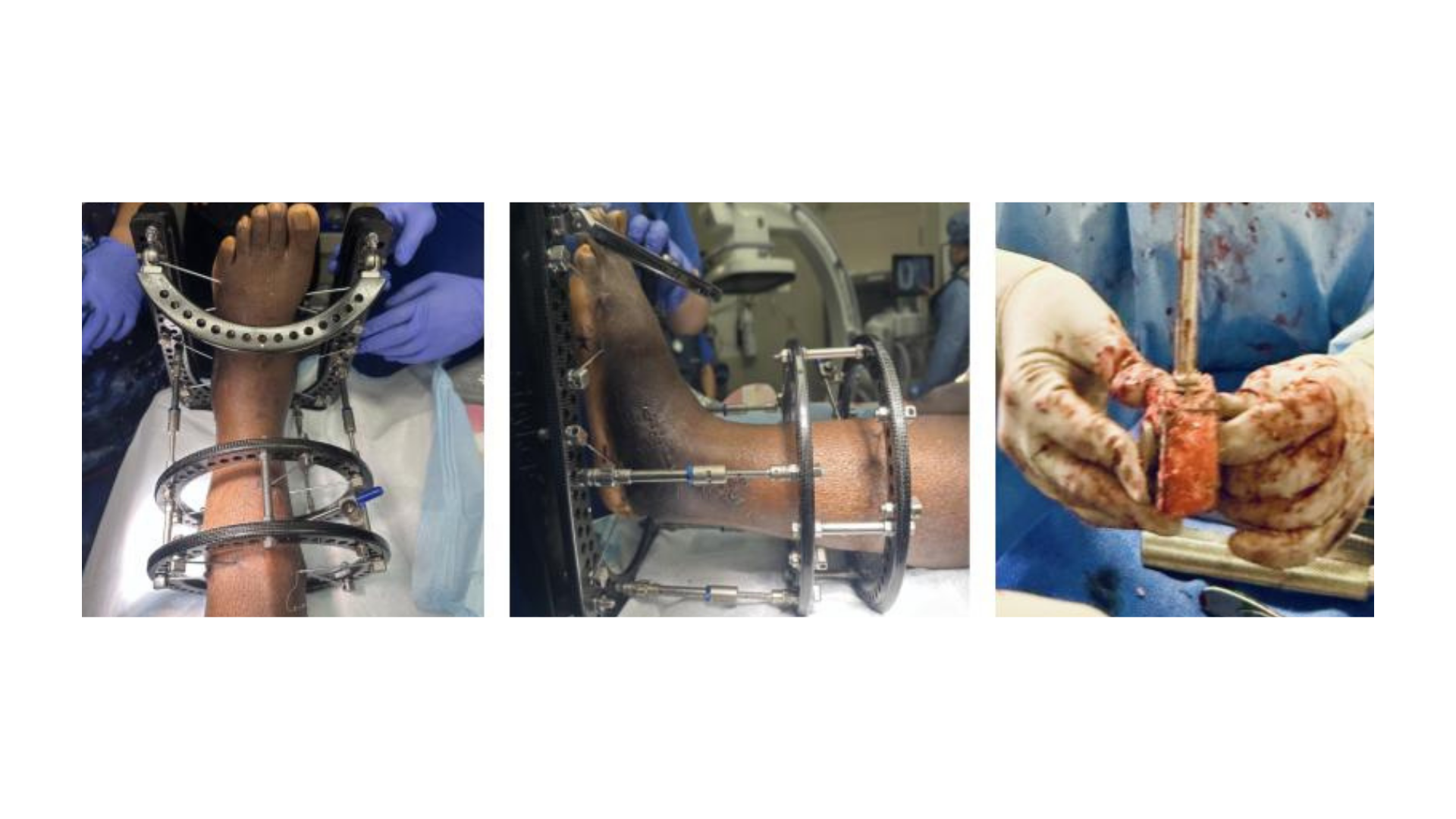
After interposition of the distal fibula between the tibia and calcaneus, we applied a circular external frame following safety zones in order to maintain alignment and allow for gradual deformity correction while protecting the soft tissues. Fluoroscopy confirmed positioning (Figure 3). Postoperatively, we instructed the patient to remain non-weight-bearing to the affected extremity until the second stage of the reconstruction. The patient was regularly monitored and had no complications before the second procedure.
Stage 2: Arthrodesis with 3D Implant (July 2025). After removing the external fixator applied in May, we undertook a definitive reconstruction. We removed the previous fibula autograft spacer and morselized it to use as graft material within the 3D cage implant (Figure 4). We noted consolidation of the resected fibula to the tibia and calcaneus, which was manually resected.
Next, we prepared, and then fenestrated, the tibial and calcaneal joint surfaces. After selecting the 3D implant based on preoperative computed tomography (CT) planning and intraoperative measurements, we placed the implant between the distal tibia and calcaneus to assist in restoring limb length and alignment. Adequate length of the extremity was noted after placement of the 3D implant.
We initiated an arthrodesis with a tibiotalocalcaneal intramedullary nail with interlocking screws. Intraoperative imaging confirmed appropriate alignment, with the foot positioned plantigrade and in slight valgus (Figure 5).
Examining the Postoperative Course
During the immediate postoperative period, the patient had been following up with us on a regular basis with no complications. The patient was non-weight-bearing for approximately 8 weeks to allow for bony consolidation, then progressed to protecting weight-bearing while initiating physical therapy.
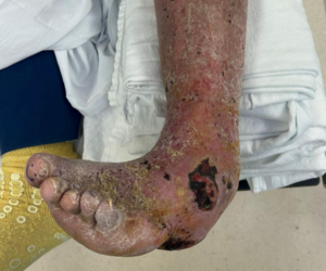
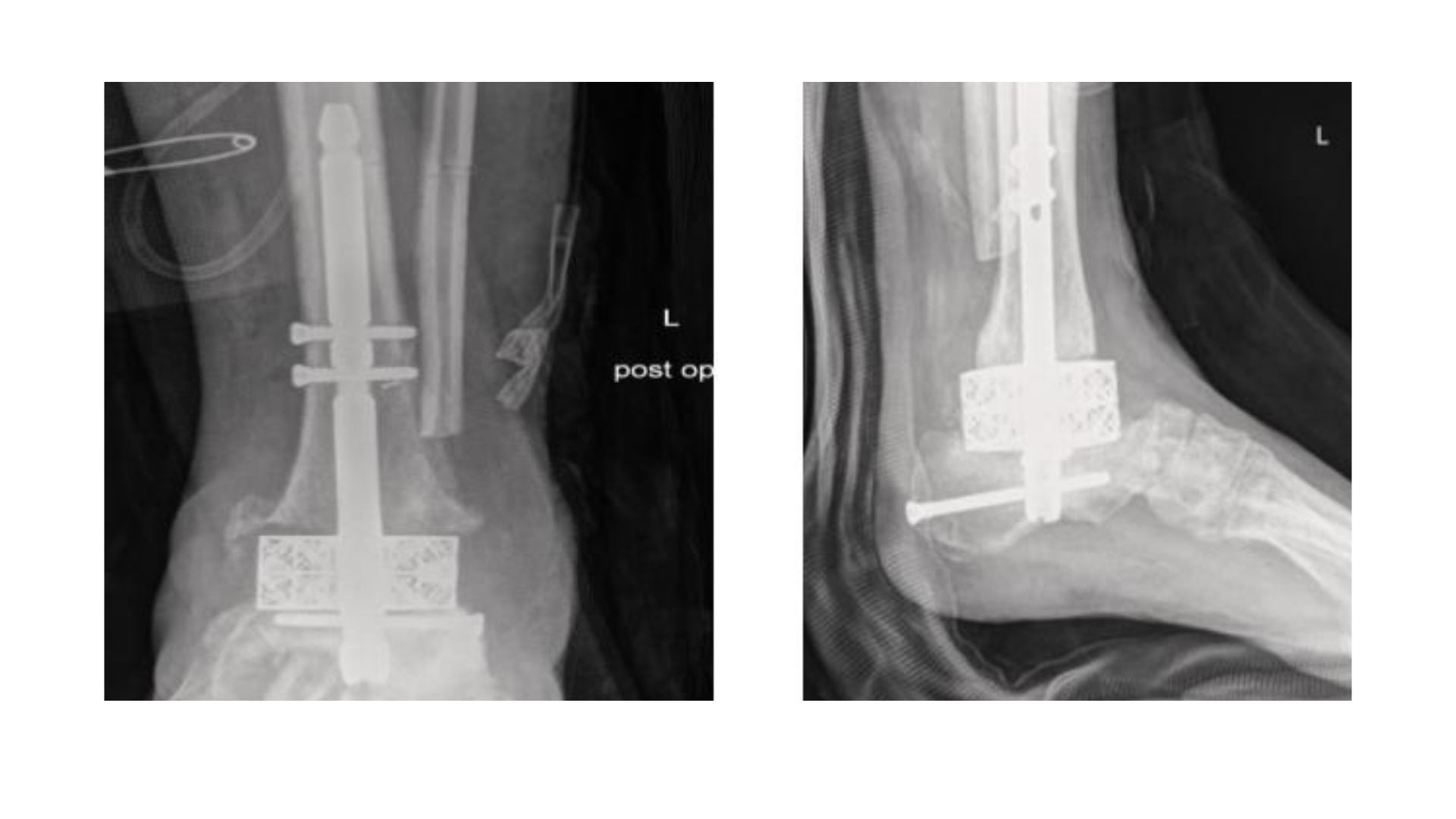
In November 2025, approximately 4 months after implant application, the patient presented with a concern of irritation to the skin and swelling. Patient was afebrile and denied any constitutional symptoms. Fluctuance was noted over the lateral aspect of the ankle and the ankle was noted to be increasingly warm and edematous. A small incision and drainage was performed in office, and deep wound cultures were obtained. We advised the patient to go to the emergency room for further workup. Laboratory evaluation was unremarkable aside from mildly elevated inflammatory markers. Imaging of the left lower extremity revealed significant perihardware lucency adjacent to the intramedullary rod at the level of the calcaneus, which we felt was suggestive of possible osteomyelitis and/or hardware loosening (Figure 6). Empiric antibiotic therapy was deferred as we planned for a prompt incision and drainage, bone biopsy, and removal of hardware.
The patient then underwent a left ankle incision and drainage with removal of hardware in the operating room. A large amount of hematoma with serosanguinous drainage was noted. A calcaneal screw was loose and floating within the heel, which we then subsequently removed. A bone biopsy and deep wound cultures were taken. Irrigation of the surgical site took place with normal saline and vancomycin. The wound was partially closed and packed with vancomycin beads. Negative pressure wound therapy was then applied over the remaining open portion of the wound. Bone biopsy, aerobic and anaerobic cultures, and blood cultures were all negative for infection. Given comorbidities, the patient completed a 6-week course of IV antibiotics as an outpatient on infectious disease’s recommendation. Postoperatively, the patient was non-weight-bearing for 4 weeks, and followed up regularly for local wound care. The wound closed during the non-weight-bearing period and the patient progressed to protected weight-bearing, continuing physical therapy.
Follow-Up Findings
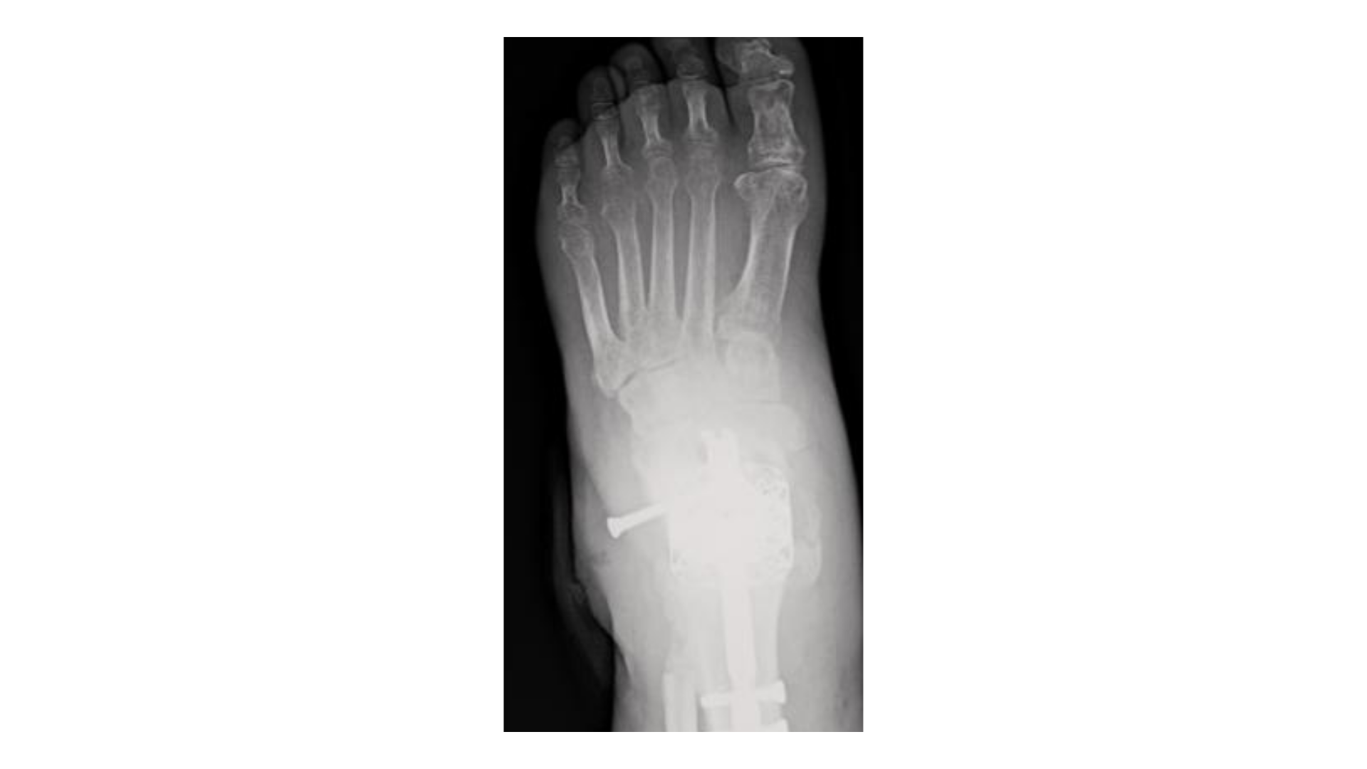
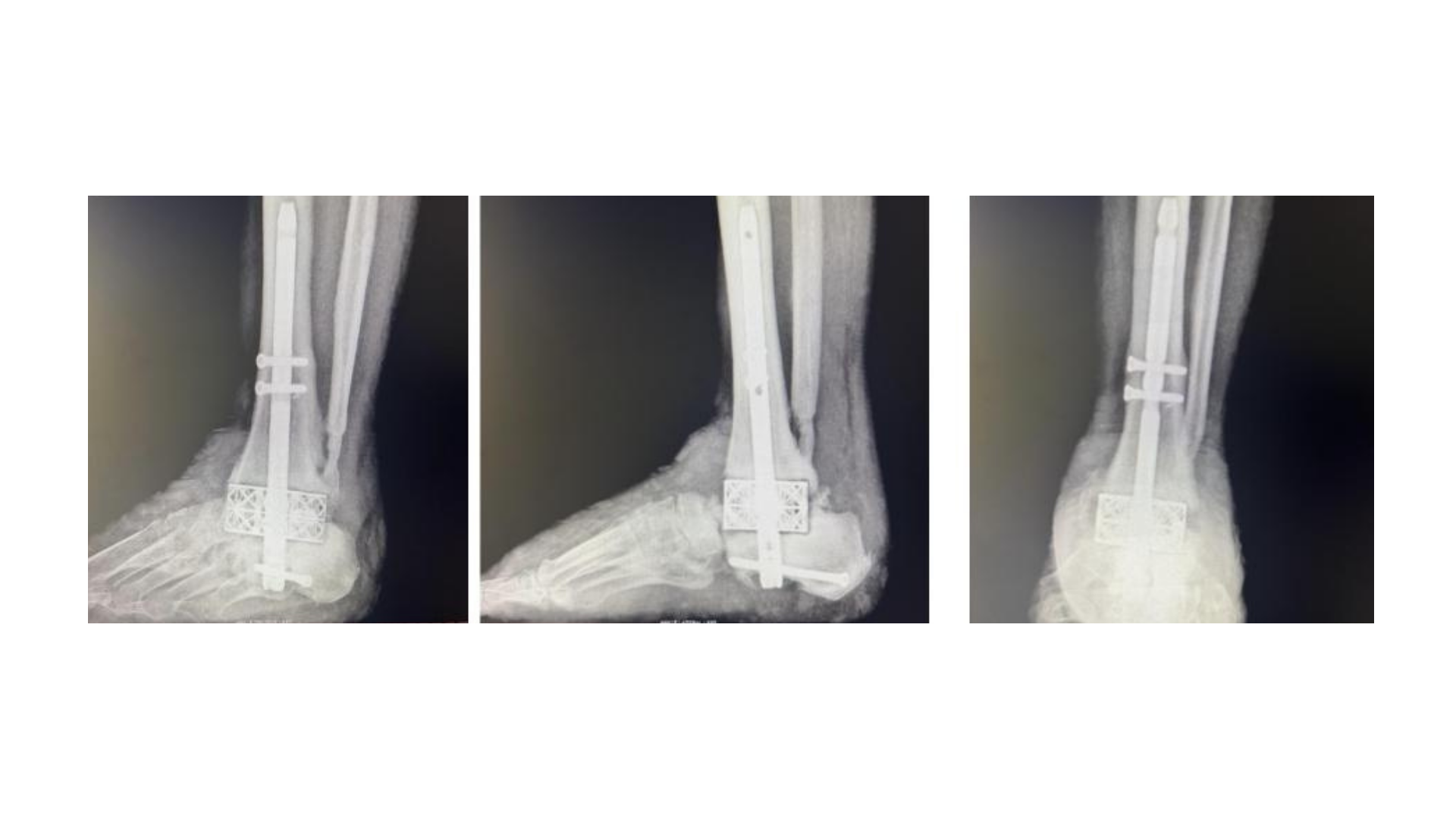
Despite complications involving hardware failure, the follow-up imaging confirms alignment and evidence of progressive fusion at the arthrodesis site at 4 months (Figure 7).
At 1-year follow-up (April 2026) the patient reported no pain during ambulation, no recurrence of ulceration or skin breakdown, independent ambulation in a CROW boot, and has returned full time to work.
Clinical evaluation and imaging confirmed a stable, plantigrade foot with successful limb salvage.
Discussion
Charcot neuroarthropathy of the ankle represents a challenging limb salvage condition. Although Charcot neuroarthropathy more commonly affects the midfoot, involvement of the ankle and hindfoot is often more difficult to manage because progressive collapse can compromise limb alignment, bracing potential, soft tissue integrity, and ambulation.8,9 The primary goals of treatment are therefore not limited to radiographic fusion, and include restoration of a stable, plantigrade, braceable extremity, prevention of recurrent ulceration, and preservation of functional ambulation.8,9
Traditional reconstruction has included arthrodesis with internal fixation, external fixation, or combined fixation constructs. However, these methods are associated with high complication rates. In a systematic review of Charcot foot reconstruction, Ha and colleagues reported an overall bone fusion rate of 86.1%. The study also reported complications directly attributable to fixation technique occurred in 36% of cases, with a post-reconstruction amputation rate of 5.5%.10 Similarly, Galhoum and team emphasized that the literature guiding ankle Charcot management is largely composed of lower-level evidence, noting that complications such as infection and loss of correction remain important concerns.9 This shows the need for adjunctive techniques that can improve structural stability defects in complex Charcot ankle reconstruction.
Large osseous defects can be challenging in ankle and hindfoot Charcot reconstruction. Bone fragmentation, talar collapse, distal tibial involvement, and loss of structural integrity can limit the ability to achieve compression and stable apposition using conventional arthrodesis techniques alone. In these cases, surgeons may consider structural grafts, external fixation, IM fixation, or custom implants. 3D printed titanium implants have emerged as a potential adjunct in these reconstructions as they can address bone loss, restore alignment, and provide a scaffold for osseous integration.11,12
In this case, the patient underwent a staged approach. The first stage involved circular external fixation, joint resection, and use of the fibula as an interpositional graft/spacer to maintain alignment. The second stage involved removal of the external fixator, preparation of the tibial and calcaneal surfaces, placement of a 3D implant, and definitive TTC arthrodesis with an IM nail. This staged approach allowed deformity correction, reassessment of the limb, and subsequent definitive reconstruction using a structural implant to address the osseous defect and restore alignment.
An important feature of this case is that the patient developed hardware related irritation, preulcerative skin changes, and radiographic evidence of hardware loosening approximately four months after definitive reconstruction. Kummen and colleagues reported that hardware failure is common following Charcot deformity correction, but that satisfactory clinical and radiographic outcomes may still be achieved in select patients despite hardware failure.13 This is consistent with our case, as the patient had an incision and drainage with hardware removal, but continued to maintain alignment and demonstrate progressive fusion. The etiology of the hardware loosening in this case remains multifactorial. The patient has a body mass index of 36 and poorly controlled diabetes mellitus. The increased BMI likely subjected the fixation construct to greater repetitive mechanical loading during the postoperative period.
The negative infectious workup in this case was also clinically significant. Given the patient’s extensive medical history and imaging findings, infection remained an important consideration. Bone biopsy, aerobic and anaerobic cultures, and blood cultures were negative. The absence of confirmed infection suggests that the complication was likely due to mechanical hardware irritation and loosening. This distinction is important because infection can threaten fusion progression and limb salvage.
Despite the complication, the patient achieved a favorable 1-year outcome. Follow up imaging demonstrated maintained alignment and progressive fusion at the arthrodesis site. Clinically, the patient reported no pain with ambulation, had no recurrent ulceration or skin breakdown, ambulated independently in a CROW boot, returned to full-time work, and maintained a stable plantigrade foot.
There are several limitations to this report. As a single case, the findings cannot be generalized to all patients with Charcot neuroarthropathy of the ankle. The patient also required a secondary procedure for hardware irritation and loosening, demonstrating that 3D printed implant technology does not eliminate the high complication burden associated with Charcot reconstruction. Additionally, 1-year follow up represents an encouraging short- to mid-term result. However, longer follow-up is necessary to evaluate durability of fusion, implant incorporation, maintenance of correction, brace dependence, recurrent ulceration, and long-term limb salvage. Larger studies are needed to better define the indications, complication rates, cost effectiveness, and long-term outcomes of 3D printed structural implants in Charcot ankle and hindfoot reconstruction.
Conclusion
This case highlights the use of 3D-printed implant within a staged reconstruction approach. Our results showed this approach can be successful in achieving limb salvage in complex Charcot ankle deformity with significant bone loss.
Although this case had screw loosening that required removal during the postoperative course, the patient maintained joint alignment and progressed to successful fusion. Ultimately, the patient achieved a pain-free, plantigrade foot, and required no assistive ambulatory device at 1-year of follow-up.
These findings can suggest that 3D implant technology can provide a successful alternative to traditional reconstruction techniques, in particular the ones involving severe osseous loss. In addition to supporting this data, other studies may contribute to evaluating long term outcomes, complication rates and cost-effective measures.
Dr. Rivera is the Chief podiatric resident at Palisades Medical Center in North Bergen, NJ. Dr. Patel is a second-year podiatric resident at Palisades Medical Center in North Bergen, NJ. Dr. Abadeer is an attending podiatrist at Palisades Medical Center and Lower Limb Institute in Hackensack, NJ.
The authors share that they have no non-financial or commercial, proprietary, or financial interest in the products or companies described in the manuscript. The author(s) did not receive grants or a consultant honorarium to conduct the study, write the manuscript or otherwise assist in the development of the above-mentioned manuscript.
References
1. van der Ven A, Chapman CB, Bowker JH. Charcot neuroarthropathy of the foot and ankle. J Am Acad Orthop Surg. 2009;17(9):562-571. doi:10.5435/00124635-200909000-00003
2. Rajbhandari S, Jenkins RC, Davies C, Tesfaye S. Charcot neuroarthropathy in diabetes mellitus. Diabetologia. 2002;45(8):1085-1096. doi:10.1007/s00125-002-0885-7
3. Pinzur MS. Surgical versus accommodative treatment for Charcot arthropathy of the midfoot. Foot Ankle Int. 2004;25(8):545-549.
4. Verity S, Sochocki M, Embil JM, Trepman E. Treatment of Charcot foot and ankle with a prefabricated removable walker brace and custom insole. Foot Ankle Surg. 2008;14(1):26-31.
5. Yammine K, Assi C. Intramedullary nail versus external fixator for ankle arthrodesis in Charcot neuroarthropathy: a meta-analysis of comparative studies. J Orthop Surg (Hong Kong). 2019;27(2):2309499019836012.
6. Rana B, Patel S. Results of ankle and hindfoot arthrodesis in diabetic Charcot neuroarthropathy—a retrospective analysis of 44 patients. J Clin Orthop Trauma. 2021;23:101637.
7. So E, Mandas VH, Hlad L. Large osseous defect reconstruction using a custom three-dimensional printed titanium truss implant. J Foot Ankle Surg. 2018;57(1):196-204.
8. Marmolejo VS, Arnold JF, Ponticello M, Andersen CA. Charcot foot: clinical clues, diagnostic strategies, and treatment principles. Am Fam Physician. 2018;97(9):594-599.
9. Galhoum AES, Trivedi V, Askar M, Tejero S, Herrera-Pérez M, Al-Rashidi Y, Valderrabano V. Management of ankle Charcot neuroarthropathy: a systematic review. J Clin Med. 2021;10(24):5923. doi:10.3390/jcm10245923
10. Ha J, Hester T, Foley R, Reichert ILH, Vas PRJ, Ahluwalia R, Kavarthapu V. Charcot foot reconstruction outcomes: a systematic review. J Clin Orthop Trauma. 2020;11(3):357-368. doi:10.1016/j.jcot.2020.03.025
11. Dekker TJ, Steele JR, Federer AE, Hamid KS, Adams SB Jr. Use of patient-specific 3D-printed titanium implants for complex foot and ankle limb salvage, deformity correction, and arthrodesis procedures. Foot Ankle Int. 2018;39(8):916-921. doi:10.1177/1071100718770133
12. Parry E, Catanzariti AR. Use of three-dimensional titanium trusses for arthrodesis procedures in foot and ankle surgery: a retrospective case series. J Foot Ankle Surg. 2021;60(4):824-833. doi:10.1053/j.jfas.2020.08.011
13. Kummen I, Phyo N, Kavarthapu V. Charcot foot reconstruction—how do hardware failure and non-union affect the clinical outcomes? Ann Joint. 2020;5:25. doi:10.21037/aoj.2020.01.06
© 2026 HMP Global. All Rights Reserved.
Any views and opinions expressed are those of the author(s) and/or participants and do not necessarily reflect the views, policy, or position of Podiatry Today or HMP Global, their employees, and affiliates.