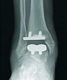
An Update On Incisional Approaches for Total Ankle Replacement
As total ankle replacement volumes rise, selecting the appropriate surgical approach remains critical to balancing implant access, deformity correction, and soft-tissue preservation. This review examines the indications, benefits, limitations, and technical considerations of anterior, anteromedial, lateral transfibular, and posterior approaches.
Key Takeaways
-
The standard anterior approach provides broad visualization and compatibility with most contemporary implant systems, but careful patient selection and meticulous soft-tissue handling are essential because wound complications remain a concern.
-
Alternative approaches offer specific advantages: the anteromedial approach may benefit selected patients with vulnerable anterior soft tissues, while the lateral transfibular approach can facilitate correction of coronal and rotational deformity.
-
The posterior approach is primarily a salvage option when anterior and lateral tissues are unsuitable; overall, incision selection should reflect vascular status, prior surgery, deformity, implant requirements, and surgeon experience.
Total ankle replacement (TAR) has evolved substantially over the past several decades, transitioning from early, high-failure designs to contemporary implants that provide reliable pain relief, preservation of ankle motion, and increasingly predictable survivorship.1-2 Due to these advancements, the rate of total ankle replacement performed also experienced substantial growth over the past 2 decades. Karzon and colleagues evaluated national trends in total ankle arthroplasty in the United States from 2009 to 2019 using a large administrative database and found a 136.1% increase in annual TAR volume, from 2,180 procedures in 2009 to 5,147 in 2019. The authors also demonstrated a 120.8% increase in population-adjusted rate.3 An abstract presented at the 2025 AOFAS Annual meeting reviewed data from the American Joint Replacement Registry demonstrating an increase in primary total ankle replacement volume from 510 procedures in 2000 to 4,893 in 2022. This represents an 859% increase in primary TAR procedures being performed. This volume was estimated to increase to 9,812 procedures in 2023 and is projected to more than double by 2040.4 This highlights the continued and substantial growth of total ankle replacement and its increasing role in the management of end-stage ankle arthritis.
Along with advances in implant design, instrumentation, and patient selection, the surgical approach used to access the ankle has remained an important and sometimes underappreciated component of total ankle arthroplasty. Historically, the standard anterior approach has been the predominant exposure for TAR.5 It provides visualization of the tibial and talar surfaces and facilitates reproducible bone preparation and component positioning without requiring osteotomy of either malleolus. The standard anterior approach is compatible with the cutting guides and instrumentation used by most contemporary TAR systems, allowing for use of surgical jigs and alignment guides without putting stress on the soft tissues.
The anterior approach still remains the gold standard due to its visualization, and broad implant compatibility; however, the anteromedial, lateral, and posterior approaches may offer important advantages in carefully selected circumstances. Ultimately, the incisional approach should be individualized based on the patient's soft tissue envelope, prior surgical history, deformity, implant system, concomitant procedures, and surgeon experience. This article reviews the anterior, anteromedial, lateral transfibular, and posterior approaches to TAR, highlighting the indications, advantages, limitations, and technical considerations that may help guide approach selection.
Preoperative Assessment
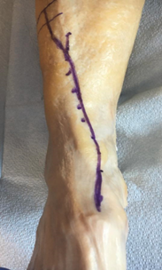
Incisional approach should begin with careful preoperative assessment of the patient’s vascular status, smoking history, diabetes status, prior surgical incisions, and the overall quality of the patient’s soft tissue envelope. The location of previous surgical incisions, skin grafts, soft tissue flaps, or areas of compromise are also considerations when determining the safest incision and surgical corridor. Surgeons should also include evaluation of the presence of associated hindfoot or midfoot pathology, as certain approaches may provide improved exposure or facilitate in adjunct procedures. Ultimately, incision selection should stem from the patient's soft tissue envelope and reconstructive needs rather than relying solely on surgeon preference.
Anterior Approach
The standard anterior incision is historically well-documented in the literature as the predominant exposure for total ankle replacement. It provides visualization of the tibial and talar surfaces and facilitates reproducible bone preparation and component positioning without requiring osteotomy of either malleolus. The standard anterior approach is compatible with the cutting guides and instrumentation used by most current TAR systems, allowing for use of surgical jigs and alignment guides without putting stress on the soft tissues.
The popularity of the anterior approach, however, should not be interpreted as evidence that it is universally appropriate. The anterior ankle incision overlies a watershed region between the anterior tibial and posterior tibial angiosomes, which has relatively poor perfusion and limited collateral overlap, predisposing to wound complications. These wound healing complications have historically represented the most common as well as detrimental complications following TAR. These concerns become particularly relevant in patients with previous anterior surgery or trauma, compromised skin, diabetes, vascular disease, inflammatory arthropathy, or other factors associated with impaired wound healing.
Raikin and team evaluated 106 total ankle arthroplasties performed through the standard anterior approach. They found that 70/106 (66%) healed uneventfully and 25% (27/106) had minor wound complications requiring local wound care and/or oral antibiotics. Eight percent (9/106) had major wound complications requiring operative treatment.6 In a matched cohort review, Reb and colleagues found delayed wound healing beyond 30 days in nearly 20% of patients undergoing either TAA or ankle arthrodesis through an anterior incision.5 Similarly, Whalen and colleagues reported a 28% rate of wound breakdown, with risk factors including smoking, peripheral vascular disease, and cardiovascular disease.6 Usuelli and team demonstrated 3.7% deep infections and (4.9%) superficial wound infections undergoing total ankle replacement using the standard anterior approach.7 Glazebrook and coworkers created an evidence based classification system for post-operative ankle arthroplasty complications.8 A retrospective review based on Glazebrook’s classification system was performed by Gadd and colleagues who found that 5.85% of 212 ankle replacements went on to have deep infection.9 Ninety percent of these patients went on to necessitate further surgical intervention and 2 patients went on to have more proximal amputations. Most recently, a study by Di Ponte in the Journal of Foot and Ankle Surgery reviewed intraoperative and postoperative complications using the standard anterior incision and the lateral transfibular approach. They found they noted wound healing problems or superficial dehiscence in 27 out of 120 standard anterior approach patients.10
Steps can be made to avoid pitfalls that can result in these potential detrimental complications. It is important to avoid excessive soft tissue retraction that can result in wound breakdown. Excessive tension from retractors can compromise both perforators and the subdermal plexus. Maintaining full thickness flaps to incorporate the anterior neurovascular bundle can avoid excessive handling. Lastly, a Doppler can be used to ensure perforators to the skin in this area are open. This is especially important in post-traumatic arthritis where the perforators can be damaged secondary to the injury.
Technique Tips
-
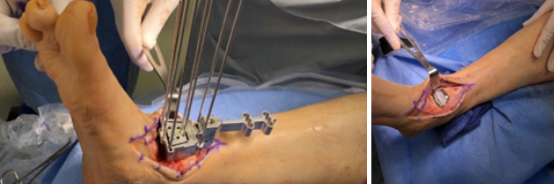
An anterior approach with a standard central incision takes place between the tibialis anterior tendon and the extensor hallucis longus tendon.
-
Care is taken to ensure full-thickness skin flaps are created medially and laterally. Maintaining the dissection in the same plane immediately deep to the subcutaneous tissue avoids unnecessary lateral or medial undermining.
-
The retinaculum is opened longitudinally in line with the skin incision.
-
An alternative retinacular incision can be made
-
Ensure there is enough retinacular tissue to facilitate anatomic closure.
-
Retract the tibialis anterior tendon medially or laterally as required to optimize visualization and accommodate the planned implant instrumentation.
-
Identify the anterior neurovascular bundle.
-
Mobilize and retract the bundle laterally, maintaining careful protection throughout the deeper dissection.
-
Identify the deep surface/floor of the tibialis anterior tendon sheath and the underlying anterior ankle capsule.
-
Perform a full-thickness capsular incision directly over the ankle joint.
-
Elevate the capsule and limited periosteal attachments from the distal tibia and talus.
-
Avoid unnecessary periosteal stripping or disruption of the surrounding soft tissues.
Anteromedial Approach
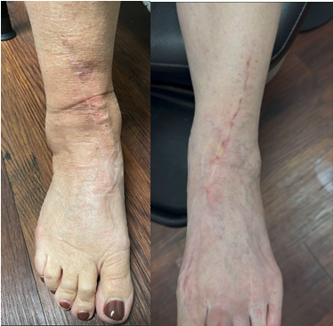
The anteromedial approach represents an alternative exposure for total ankle arthroplasty that was developed primarily in response to the significant wound-healing complications associated with the standard anterior approach. The rationale for this technique is based on angiosome anatomy, with the incision positioned at the interface between the anterior tibial and posterior tibial angiosomes rather than directly through the central anterior ankle skin. Disruption of the angiosomes of the ankle not only increases the likelihood of incisional dehiscence, but also makes coverage via adipofacial or musculocutaneous flaps difficult.8 By maintaining the vascular territories and associated perforating vessels, this approach may preserve a more favorable blood supply to the skin flaps and potentially decrease the risk of postoperative wound complications.
In 2013, Bibbo first described a modification to the anterior approach that preserves the remaining skin perforator vessels and prevents intertissue plane shearing of fragile skin and subcutaneous tissue. The incision is initiated along the distal third of the leg and directed toward the anteromedial ankle remaining just lateral to the tibialis anterior tendon sheath. It is continued distally along the medial aspect of the ankle toward the transverse tarsal joint before being redirected toward the midline and extended distally as necessary to achieve adequate, tension-free soft-tissue mobilization.11 Rodriguez and colleagues later advanced the concept by introducing an orthoplastic anteromedial approach. This incision lies fully within the angiosome of the anterior tibial artery, medial to the central watershed zone, thereby respecting the vascular integrity of the region. Rodriguez-Collazo and team retrospectively evaluated 27 total ankle arthroplasties performed through an angiosome-based anteromedial approach and reported a wound complication in 1 patient (3.7%).12 More recent data has further emphasized the importance of preoperative vascular assessment when considering an anteromedial approach. LaPorta and colleagues retrospectively evaluated 23 patients undergoing either total ankle or total talus replacement through an orthoplastic anteromedial approach. Eighteen patients healed uneventfully, while the authors emphasized that assessment of the vascular supply is necessary to determine the healing potential of the approach. This is particularly relevant because the theoretical benefit of the anteromedial incision is dependent on preservation of adequate perfusion within the adjacent angiosomes.13
Despite these potential advantages, there are several limitations that should be recognized. All studies are retrospective in nature with relatively small cohorts. Differences in patient selection, surgeon experience, implant design, perioperative wound management, and the timing of adoption of the approach introduce potential limitations. Another important limitation is compatibility with modern total ankle arthroplasty systems and their instrumentation. Most total ankle systems were designed around an anterior surgical approach. Their associated cutting guides, alignment jigs, and instrumentation may be more accommodative through the standard anterior approach. The anteromedial approach may require modification of the surgical technique or instrumentation depending on the implant system being utilized. If the incision is placed too medial this can result in the inability to utilize instrumentation associated with the replacement. Limited lateral exposure may also make management of lateral gutter pathology more challenging. Lastly, to the authors’ knowledge, no study has directly compared wound healing complications between the standard anterior and anteromedial approaches within a single cohort.
Although limited, the anteromedial approach represents a promising alternative to the traditional standard anterior incision for total ankle arthroplasty, particularly in patients with compromised anterior soft-tissue envelopes or elevated risk for wound-healing complications.
Technique Overview
-
Identify and mark the tibial crest proximally.
-
Begin the incision approximately 1 cm lateral to the tibial crest.
-
At approximately 5 cm proximal to the ankle joint, transition medially toward the tibialis anterior tendon as the anterior tibia becomes flatter.
-
Continue the incision along the medial border of the tibialis anterior tendon, curving medially as it progresses distally.
-
Extend distally as needed to obtain adequate, tension-free exposure of the ankle joint.
-
Identify the tibialis anterior tendon and its sheath.
-
Open the tendon sheath longitudinally to facilitate mobilization of the tendon.
-
Retract the tibialis anterior tendon medially or laterally as required to optimize visualization and accommodate the planned implant instrumentation.
-
Identify the anterior neurovascular bundle.
-
Mobilize and retract the bundle laterally, maintaining careful protection throughout the deeper dissection.
-
Identify the deep surface/floor of the tibialis anterior tendon sheath and the underlying anterior ankle capsule.
-
Perform a full-thickness capsular incision directly over the ankle joint.
-
Elevate the capsule and limited periosteal attachments from the distal tibia and talus.
-
Avoid unnecessary periosteal stripping or disruption of the surrounding soft tissues.
Lateral Transfibular Approach
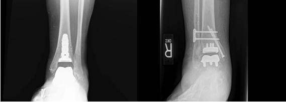
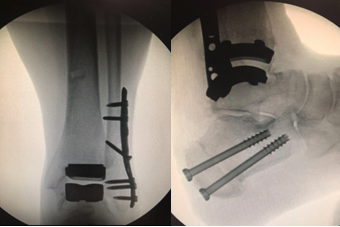
The lateral transfibular approach was initially introduced in part because avoidance of the anterior ankle incision was hypothesized to reduce wound healing complications by preserving the anterior soft tissue envelope. However, subsequent clinical data have not consistently demonstrated a lower incidence of wound complications compared with the anterior approach. The lateral transfibular approach represents a relatively recent evolution in total ankle arthroplasty, in our observation, gaining prominence with the development of the Zimmer Trabecular Metal Total Ankle Replacement in the early 2010s. Unlike the traditional anterior approach, the transfibular technique provides direct lateral access to the tibiotalar joint through a fibular osteotomy, allowing improved visualization of the ankle center of rotation and facilitating anatomically curved resections of the tibial plafond and talus. The approach was developed in conjunction with implant designs specifically intended for lateral access, with the goal of minimizing bone resection, reproducing native joint geometry, and allowing correction of coronal, sagittal, and rotational deformity, including deformity related to fibular malposition or shortening.14
The primary advantage is the extensile lateral exposure and ability to directly address coronal plane deformity and fibular length while avoiding an anterior incision and its associated soft tissue envelope. However, these benefits must be balanced against the morbidity of a fibular osteotomy, which requires reliable reduction and fixation and introduces risks of delayed union, nonunion, malunion, syndesmotic instability, hardware irritation, and potential hardware removal. The approach may also make ligamentous balancing and polyethylene sizing more technically demanding, and its use is generally dependent on an implant specifically designed for the transfibular corridor.
More recent literature suggests that the lateral approach does not necessarily show a significant reduction in wound complications compared with modern anterior approaches; rather, its primary contemporary advantage appears to be deformity correction and improved lateral visualization. Despite the additional osseous work, published series demonstrate encouraging mid- and long-term results, with a systematic review reporting a fibular nonunion rate of approximately 1% among total ankle arthroplasty cases and recent ≥5-year series demonstrating durable alignment and implant survival. Gardini and colleagues performed a meta-analysis of 51 studies and 7,959 TARs found wound complications in 2.32% of lateral/transfibular cases (17/734) versus 1.83% with anterior approaches (132/7,225).This difference was not statistically significant (P=0.319). Usuelli and coworkers published a comparative series of 150 TARs found superficial infection rates of 2.9% with the lateral approach vs 4.9% with the anterior approach, while deep infection occurred in 1.4% vs 3.7%, respectively.15-16 From the data noted above, the main advantage of the transfibular approach appears to be its direct lateral exposure, ability to address coronal and rotational deformity, and compatibility with implant designs rather than the reduction in wound morbidity.
Technique Overview
-
Patient placement is in the supine position with a bump beneath the ipsilateral hip to internally rotate the extremity
-
A longitudinal incision centers over the distal fibula and extends proximally and distally as necessary.
-
The surgeon carries the incision through the skin and subcutaneous tissue with meticulous preservation of the soft tissue envelope.
-
The superficial peroneal is identified and protected, particularly with more proximal extension of the incision.
-
The subperiosteal exposure of the distal fibula takes place at the level planned for the osteotomy.
-
One determines the level and orientation of the osteotomy based on the implant system and desired exposure.
-
An oblique fibular osteotomy is then performed
-
A transverse or chevron osteotomy can also be utilized.
-
The osteotomy should provide adequate access to the lateral ankle while preserving sufficient proximal and distal fibular bone for stable fixation.
-
The fibular segment is mobilized and reflected posteriorly and inferiorly
-
Care must be taken to preserve the syndesmosis and lateral soft tissue
-
The anterior talofibular and calcaneofibular ligamentous structures may require release to obtain adequate exposure and facilitate correction.
-
The distal tibiofibular and lateral capsular structures are released according to the requirements of the deformity and implant system.
-
The ankle capsule is incised and elevated to expose the tibiotalar joint.
Posterior Approach
In cases when the anterior and lateral soft tissue envelopes are compromised and total ankle replacement is still desired the posterior approach can be utilized as a last-ditch effort. In 2013, Bibbo first described use of the posterior approach in a 44-year-old male with history of an open tibial fracture 8 years prior. This resulted in significant scarring of the anterior soft tissue envelope, recurvatum deformity of the distal tibia, equinus deformity, and finally end stage ankle arthrosis. A staged approach was performed with correction of the tibial deformity. The second stage was performed utilizing direct posterior approach with Z-lengthening and retraction of the Achilles tendon, lengthening of the flexor hallucis longus, and retraction of the remaining posterior structures to expose the ankle joint, allowing correction of residual supramalleolar varus and posterior insertion of a stemmed total ankle prosthesis. At the 2-year follow-up, the patient was ambulating without pain in normal shoe gear with a final ROM of an arc of 7 degrees, with a range of 4 degrees of plantarflexion to 3 degrees of dorsiflexion.17
A poster at the ACFAS Annual Scientific Conference by Foote and colleagues described the use of the posterior approach in a 43 year old male with a history of an open talar fracture dislocation, calcaneal fracture, resulting in extensive anterolateral soft tissue loss. This ultimately required a radial forearm free-flap for coverage. Following treatment of chronic talar osteomyelitis with debridement and antibiotic cement spacer, the patient underwent a staged total ankle replacement through a posterior approach. At 1-year follow up, the patient had maintained anatomic alignment. Functional outcomes were assessed using the American Orthopaedic Foot & Ankle Society (AOFAS) hindfoot score and Lower Extremity Functional Scale (LEFS). Preoperatively, the AOFAS and LEFS scores were 9 and 15% respectively, improving to 81 and 77% at the 1-year postoperative follow-up.18
While the anterior and lateral approaches remain more commonly utilized in total ankle replacement, the posterior approach is a rarely discussed alternative that may offer distinct advantages in patients with specific anatomic, deformity, or soft tissue considerations. The posterior approach offers several potential advantages including direct access to the posterior ankle joint, with excellent visualization of the posterior tibial plafond and talus, the ability to address posterior osteophytes, posterior capsule contracture, equinus contracture, as well as a robust soft tissue envelope for coverage. However, this approach as several disadvantages including limited implant options, technically difficult with challenges of component positioning and instrumentation, potential injuries to the sural nerve and posterior tibial neurovascular bundle. There is also an additional concern of poor wound healing overlying the Achilles tendon. At this time the posterior approach should be utilized in salvage cases and those in which the anterior and lateral soft tissue envelopes are not amenable for
Posterior Technique Overview
-
The patient is placed in the prone position with appropriate padding of all bony prominences.
-
Ensure the operative leg can be manipulated freely and that fluoroscopy can obtain AP, lateral, and mortise views.
-
A longitudinal incision centers over the Achilles tendon.
-
The incision should provide adequate proximal and distal exposure while avoiding excessive extension that could compromise the soft tissue envelope.
-
Incise the skin and carefully dissect through the subcutaneous tissues to identify the paratenon.
-
Care is taken to preserve the paratenon and surrounding soft tissue attachments when possible.
-
Incise the Achilles paratenon longitudinally and expose the tendon.
-
The Achilles may be split longitudinally or lengthened using a Z-plasty to facilitate access to the posterior ankle.
-
Dissection is carried deep to access the deep posterior compartment.
-
Identify and protect the posterior tibial neurovascular bundle, which lies anterior to the flexor hallucis longus region.
-
The posterior ankle capsule is identified and a longitudinal posterior capsulotomy is performed to expose the posterior aspect of the tibiotalar joint.
-
Finally, the capsule is elevated as necessary to visualize the posterior tibial plafond and talar dome.
Adjunctive Strategies Across Approaches
Several adjuncts can be utilized to optimize soft-tissue healing following total ankle replacement (TAR). Current evidence does not support routine use of any single intervention. Tranexamic acid (TXA), administered intravenously or topically, may reduce perioperative bleeding and hematoma formation, with both routes demonstrating utility in lower-extremity arthroplasty; however, TAR-specific evidence remains limited. Similarly, closed-incision negative-pressure wound therapy (ciNPWT) has been advocated to reduce edema, wound drainage, and dehiscence, particularly in patients with compromised soft-tissue envelopes. Small retrospective series have reported favorable wound healing rates with ciNPWT following anterior TAR, including successful healing in high-risk patients, although these findings have not been consistently reproduced in comparative studies. In their systematic review of 29 studies consisting of 6,986 TARs, Sakkab and colleagues found that comparisons of conventional closure with adjunctive strategies including negative-pressure wound therapy and TXA did not demonstrate statistically significant differences in wound outcomes.19 These findings suggest that such adjuncts may be most appropriately reserved for selected high-risk patients rather than considered substitutes for meticulous surgical technique and appropriate patient selection. Meticulous soft tissue handling remains the most important component of incision management during TAR.
Conclusion
Total ankle replacement has experienced substantial growth with continued advances in implant design, instrumentation, and patient selection. Although the standard anterior approach remains the most widely utilized exposure because of its visualization and compatibility with contemporary instrumentation, concerns regarding wound-healing complications have prompted increasing interest in alternative approaches. This article reviews the anterior, anteromedial, lateral transfibular, and posterior approaches to total ankle replacement, emphasizing the importance of individualized approach selection based on the patient's soft-tissue envelope, vascular status, prior surgical history, deformity, concomitant pathology, implant system, and surgeon experience. The anteromedial approach may offer a soft tissue advantage in appropriately selected patients by respecting angiosome anatomy, while the lateral transfibular approach provides excellent lateral visualization and facilitates correction of coronal and rotational deformity, albeit with the added morbidity of fibular osteotomy. The posterior approach remains a rarely utilized salvage option for patients in whom the anterior and lateral soft-tissue envelopes are unsuitable, with potential advantages in exposure and soft-tissue coverage but significant technical limitations and restricted implant compatibility. Across all approaches, meticulous soft-tissue handling, appropriate patient selection, and consideration of adjunctive wound-management strategies remain essential. Ultimately, no single incision is universally optimal, and approach selection should be individualized to balance surgical exposure, deformity correction, implant requirements, and preservation of the soft-tissue envelope.
Dr. Spingola is a fellowship-trained foot and ankle surgeon and a Fellow of the American College of Foot and Ankle Surgeons, practicing in Oklahoma City, OK.
Dr. Le is a fellowship-trained foot and ankle surgeon and a Fellow of the American College of Foot and Ankle Surgeons, practicing in Montreal, Quebec.
References
-
Pugely AJ, Lu X, Amendola A, Callaghan JJ, Martin CT, Cram P. Trends in the use of total ankle replacement and ankle arthrodesis in the United States Medicare population. Foot Ankle Int. 2014;35(3):220-226. doi:10.1177/1071100713511606.
-
Zaidi R, Cro S, Gurusamy K, et al. The outcome of total ankle replacement: a systematic review and meta-analysis. J Bone Joint Surg Br. 2013;95-B(11):1500-1507. doi:10.1302/0301-620X.95B11.31633.
-
Karzon AL, Kadakia RJ, Coleman MM, Bariteau JT, Labib SA. The Rise of Total Ankle Arthroplasty Use: A Database Analysis Describing Case Volumes and Incidence Trends in the United States Between 2009 and 2019. Foot Ankle Int. 2022;43(11):1501-1510. doi:10.1177/10711007221119148.
-
Zak SG, et al. Projections and Epidemiology of Total Ankle and Revision Total Ankle Arthroplasty in the United States to 2030. Foot Ankle Spec. The National Inpatient Sample analysis found a 564% increase in TAA from 2005 to 2017.
-
Reb CW, Watson BC, Fidler CM, Van Dyke B, Hyer CF, Berlet GC, Prissel MA. Anterior ankle incision wound complications between total ankle replacement and ankle arthrodesis: a matched cohort study. J Foot Ankle Surg. 2021;60(1):47-50. doi:10.1053/j.jfas.2020.04.015.
-
Raikin SM, Kane J, Ciminiello ME. Risk factors for incision-healing complications following total ankle arthroplasty. J Bone Joint Surg Am. 2010;92(12):2150-2155. doi:10.2106/JBJS.I.00870.
-
Usuelli FG, Indino C, Maccario C, Manzi L, Liuni FM, Vulcano E. Infections in primary total ankle replacement: anterior approach versus lateral transfibular approach. Foot Ankle Surg. 2019;25(1):19-23. doi:10.1016/j.fas.2018.01.001.
-
Glazebrook MA, Arsenault K, Dunbar M. Evidence-based classification of complications in total ankle arthroplasty. Foot Ankle Int. 2009;30(10):945-949. doi:10.3113/FAI.2009.0945.
-
Gadd RJ, Barwick TW, Paling E, Davies MB, Blundell CM. Assessment of a three-grade classification of complications in total ankle replacement. Foot Ankle Int. 2014;35(5):434-441. doi:10.1177/1071100714524549.
-
Di Ponte M, Caravelli S, Viotto M, Cassanelli E, Di Maio L, Di Pasquale G, Baiardi A, Berveglieri L, Gardini G, Capodagli C, Mosca M. Comparison of anterior and lateral approaches for total ankle replacement: a retrospective cohort study. J Foot Ankle Surg. 2026. doi:10.1053/j.jfas.2026.05.017.
-
Bibbo C. A modified anterior approach to the ankle. J Foot Ankle Surg. 2013;52(1):136-137. doi:10.1053/j.jfas.2012.10.010.
-
Rodriguez-Collazo E, Agyen J. Low incidence complication with anteromedial angiosome based approach to total ankle arthroplasty: a retrospective analysis of 27 cases. Clin Surg. 2018;3:2000. doi:10.25107/2474-1647.2000
-
LaPorta GA, Schnack LL, Begum M, Yates EA, Oexeman S, Rodriguez-Collazo ER. A retrospective review of soft-tissue complications in total talus replacement and total ankle replacement using the orthoplastic anteromedial approach. J Am Podiatr Med Assoc. 2025;115(4):23-090. doi:10.7547/23-090.
-
Usuelli FG, Indino C, Maccario C, Manzi L, Salini V. Total ankle replacement through a lateral approach: surgical tips. SICOT J. 2016;2:38. doi:10.1051/sicotj/2016029.
-
Gardini F, et al. Different complication patterns between anterior and lateral approaches in total ankle arthroplasty: a systematic review and meta-analysis. J Exp Orthop. 2026. doi:10.1002/jeo2.70774.
-
Barg A, Bettin CC, Burstein AH, Saltzman CL, Gililland J. Early clinical and radiographic outcomes of trabecular metal total ankle replacement using a transfibular approach. Foot Ankle Int. 2013;34(10):1310-1321. doi:10.1177/1071100713492132.
-
Bibbo C. Posterior approach for total ankle arthroplasty. J Foot Ankle Surg. 2013;52(1):132-135. doi:10.1053/j.jfas.2012.10.007.
-
Foote GA, Bala VA, Keeley CR, Piraino J. Posterior approach for total ankle replacement. Presented at: American College of Foot and Ankle Surgeons Annual Scientific Conference; 2020.
-
Sakkab R, Radcliffe MJ, Paramasivam D, McAlister JE. Total ankle arthroplasty incision management: What Matters? A systematic review. Foot & Ankle Surgery: Techniques, Reports & Cases. 2024;4(4):100433.
© 2026 HMP Global. All Rights Reserved.
Any views and opinions expressed are those of the author(s) and/or participants and do not necessarily reflect the views, policy, or position of Podiatry Today or HMP Global, their employees, and affiliates.