Does COVID-19 Influence Postoperative Deep Vein Thrombosis?
Citing the known association of COVID-19 infection with hypercoagulability and a prothrombotic state, a team of clinicians studied the incidence of deep vein thrombosis (DVT) after foot and ankle surgery in the context of a chronologically related COVID-19 diagnosis.1
Did a COVID-19 infection in relative proximity to foot and ankle surgery have an impact on the incidence of DVT? And, did vaccination status play a role at all?
Researchers analyzed patient records from March 23, 2020 to July 31, 2022 of adults having foot and ankle surgery at one institution. They presented their results in a poster at the American College of Foot and Ankle Surgeons (ACFAS) Annual Scientific Conference. Each patient in the cohort also had a documented COVID-19 infection sometime during the study period, even if not directly during the operative timeframe. They then stratified those infections in relation to their procedure as either “recent” (10-120 days before surgery), “current” (less than 10 days preoperatively), or “postoperative” (less than 30 days after surgery).1
Any other patients studied who did not meet the above timeframes then became the “no COVID” perioperative cohort. Researchers then gathered data on these patients, including medical history, surgery type, timing of COVID-19 infection, and incidence of DVT.1 A total of 472 patients were included, with 321 having a perioperative COVID-19 infection by the researchers’ definition. Symptomatic DVT among those having foot and ankle surgery and a perioperative COVID-19 diagnosis took place at a rate of 3.12%. The DVT rate among the “no COVID” group was 1.99%, which was not found to be statistically significant. They additionally broke the “recent” COVID-19 cohort into monthly subcohorts, and found no change in the data conclusion.
Looking at vaccination status, the authors found no differences in DVT rate among unvaccinated, partially vaccinated, and fully vaccinated individuals.1 In the setting of a COVID-19 diagnosis, the anatomic location or trauma reason of the surgery also did not reveal an impact on the DVT incidence in this group. Although previous DVT and history of coronary artery disease did show a postoperative correlation with DVT incidence, there was no evidence that COVID-19 contributed to these increased rates.
Recognizing limitations of human error in review, DVTs diagnosed outside of the study facility, and nuances of false positives/negatives in COVID-19 testing, the authors concluded that from this data set, the findings suggest that COVID-19 infection in the perioperative period does not increase symptomatic rates of DVT after foot and ankle surgery.1
Reference
1. Bischoff A, Joseph N, Roby M, Judickas S, Barron I. The influence of COVID-19 on the rate of symptomatic deep vein thrombosis following foot and ankle surgery. Poster presented at the American College of Foot and Ankle Surgeons Annual Scientific Conference. February 1-3, 2024. Tampa, FL.
Hindfoot Alignment Assessment Prior to Midfoot Charcot Reconstruction: Assessing Risks
How could hindfoot alignment impact outcomes in midfoot Charcot reconstruction? Authors of a recently presented poster at the American College of Foot and Ankle Surgeons Annual Scientific Conference took on this question, looking 210 patients treated over a 17-year period.1
Records were retrospectively reviewed for these patients with midfoot Charcot neuroarthropathy who underwent reconstruction. Researchers analyzed preoperative hindfoot alignment using the Saltzman radiographic view, classifying the result as either valgus, neutral, or varus.1 Then the authors examined postoperative outcomes, specifically data associated with new osteomyelitis site, new Charcot site, hardware failure/breakage, and pin tract infection.
The authors found that both varus and valgus hindfoot alignment had more complications after reconstruction than those with neutral alignment.1 They contended that such malalignment may contribute to these challenges. Specifically, compared to neutral hindfoot alignment, new Charcot collapse occurred 4.9 times more often, pin tract infection 5.8 times more often, and hardware failure 3.5 times more often. Looking at varus-aligned hindfeet, pin tract infections carried 5.8 times higher odds, and osteomyelitis 2.8 times higher odds than patients with neutral hindfeet. Additionally, they found that valgus alignment in this cohort carried a 5.3-times higher incidence of new osteomyelitis than those patients in varus.
Stressing the importance of evaluating hindfoot alignment as part of preoperative planning for midfoot Charcot reconstruction, the authors feel these insights may aid in enhanced risk assessment, and informed surgical decision-making in these complex cases.1
Reference
1. Parkman L, Milisits T, Tefera E, et al. Hindfoot alignment effect on midfoot Charcot reconstruction. Poster presented at the American College of Foot and Ankle Surgeons Annual Scientific Conference. February 1-3, 2024. Tampa, FL.
Could a New Technique in Transmetatarsal Amputation Lead to Better Outcomes?
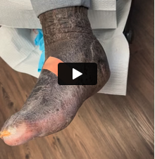
Surgeon authors of a recent poster from the American College of Foot and Ankle Surgeons Annual Scientific Conference looked at the results from a novel closure technique for transmetatarsal amputation in patients with and without peripheral vascular disease in a retrospective comparative study (49 feet, 47 patients).1 One group of patients underwent vascular intervention for peripheral vascular disease (Group 1), and one group did not (Group 2).
They described the novel components of the surgical technique as dissection of the “forefoot off the plantar flap using Terashi’s method2 for preserving intermetatarsal soft tissue.”1 After flushing the area, layered closure was first with 2-0 braided absorbable suture to affix the plantar muscle over the bone stumps, and then closure of deep and subcutaneous layers. Non-absorbable braided suture was used in skin closure for these cases.1 No patients had concomitant Achilles tendon lengthening and any cases not fully closeable primarily (49%) had standard wound care and advanced interventions as needed. Intravenous antibiotic decisions came from infectious disease, and revascularization need was determined by vascular surgery.
Among those studied, 50% of Group 1 healed, in comparison to 72% in Group 2.1 At one year, 35% of Group 1 remained healed and 66% in Group 2. Below- or above-knee amputation was an end result in 35% of Group 1 and in 6% of Group 2. The authors contend that these outcomes reveal better healing rates than those found in their cited literature review, suggesting that this method may be promising, especially for patients without vascular compromise. They also noted that absorbable suture did not seem to reveal any negative impact.1
References
1. Ennis H, Chiu H. Outcomes of transmetatarsal amputation with a modified technique utilizing muscle for closure. Poster presented at the American College of Foot and Ankle Surgeons Annual Scientific Conference. February 1-3, 2024. Tampa, FL.
2. Terashi H, Kitano I, Tsuji T, Hashikawa K, Tahara S. A modified transmetatarsal amputation. J Foot Ankle Surg. 2011;50(4):441-444.
Do First Metatarsophalangeal Joint Fusion Rates Vary With Fixation Method?
Fixation pathways vary among surgeons for first MTPJ fusion, so the authors of a poster presented at the American College of Foot and Ankle Surgeons Annual Scientific Conference aimed to examine 3 different methods to assess any differences in arthrodesis achievement. They looked at 147 patients who underwent this procedure with 13 surgeons over a more than 4-year period with the following fixation methods1:
- Dorsal plate;
- Dorsal plate with interfragmentary screw; or
- Interfragmentary screws only.
They assessed achievement of fusion from radiographs. On average the plate-only group fused in 9.02 weeks, the plate and screw group in 12.5 weeks and screws only group in 9.8 weeks.1 No significant statistical difference between the groups was appreciated by the researchers (P = .325).1 The authors noted only 4% of the cohort underwent screw-only fixation, and that other comorbidities are important, such as weight-bearing protocols and comorbidities. Surgical techniques, they stated, could also vary among surgeons, as may radiographic protocols and timing. They recommend further study with a larger cohort and incorporating these additional factors into the data.1
Reference
1. Mosseri A, Hlad LM. A retrospective analysis of fixation methods for first MPJ arthrodesis: a comparative study on fusion rates in 147 patients. Poster presented at the American College of Foot and Ankle Surgeons Annual Scientific Conference. February 1-3, 2024. Tampa, FL.