

Ballerina Foot in Heart
© 2024 HMP Global. All Rights Reserved.
Any views and opinions expressed are those of the author(s) and/or participants and do not necessarily reflect the views, policy, or position of the Journal of Invasive Cardiology or HMP Global, their employees, and affiliates.
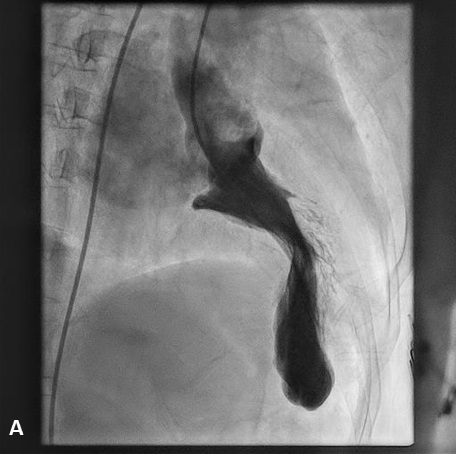
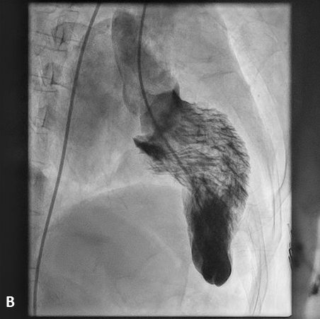
A 67-year-old woman with no known risk factors for coronary artery disease presented to the outpatient department with complaints of exertional fatigue and angina New York Heart Association class III. A 12-lead electrocardiogram showed sinus rhythm, left atrial enlargement, and left ventricular hypertrophy. A 2-dimensional echocardiogram revealed asymmetrical septal hypertrophy, left ventricular outflow tract obstruction (LVOTO) with a gradient of 124 mm Hg, mitral annular calcification (MAC), and moderate mitral regurgitation (MR).
After obtaining informed consent, the patient was taken up for a left heart catheterization and coronary angiography to look for an appropriate septal branch for alcohol septal ablation. The left ventricular systolic pressure was significantly elevated (LVESP 250 mm Hg) and the left ventricle (LV)-to-aorta pullback showed an intracavitary gradient of 140 mm Hg, with no left ventricular outflow tract (LVOT)-to-aorta gradient. The LV angiogram showed the “ballerina foot deformity”, which represents the asymmetrical septal hypertrophy with LV hypercontractility; and severe mitral annular calcification with grade 3 mitral regurgitation (Figure, Videos 1 and 2). Her coronary angiography revealed a significant obstructive disease in the mid-left anterior descending artery (LAD), while the rest of the vessels were normal. After discussion with the heart team, she was taken up for alcohol septal ablation, which successfully decreased the LVOTO gradient to 25 mm Hg. Subsequently, percutaneous coronary angioplasty to the LAD was done successfully and the patient was discharged with significant symptomatic relief.
Ballerina foot appearance is a long-described LV angiography sign in hypertrophic obstructive cardiomyopathy that reflects LV hypercontractility with a resultant narrow LV systolic cavity, resembling a ballet dancer’s feet. The eponym has also been used to describe the appearance of basal LV hypercontractility and the visibly prolapsing leaflet on LV angiography in mitral valve prolapse syndrome.
Affiliations and Disclosures
From the Department of Cardiology, All India Institute of Medical Sciences, New Delhi, India.
Disclosures: The authors report no financial relationships or conflicts of interest regarding the content herein.
Address for correspondence: Sourabh Agstam, MD, DM, MRCP(UK), FACC, Department of Cardiology, All India Institute of Medical Sciences, New Delhi, India. Email: sourabhagstam@gmail.com; X: @agstamsourabh
Back to September 2024 Table of Contents


