

Multiple Plaque Ruptures Healing Assessed by Serial Optical Coherence Tomography
© 2024 HMP Global. All Rights Reserved.
Any views and opinions expressed are those of the author(s) and/or participants and do not necessarily reflect the views, policy, or position of the Journal of Invasive Cardiology or HMP Global, their employees, and affiliates.
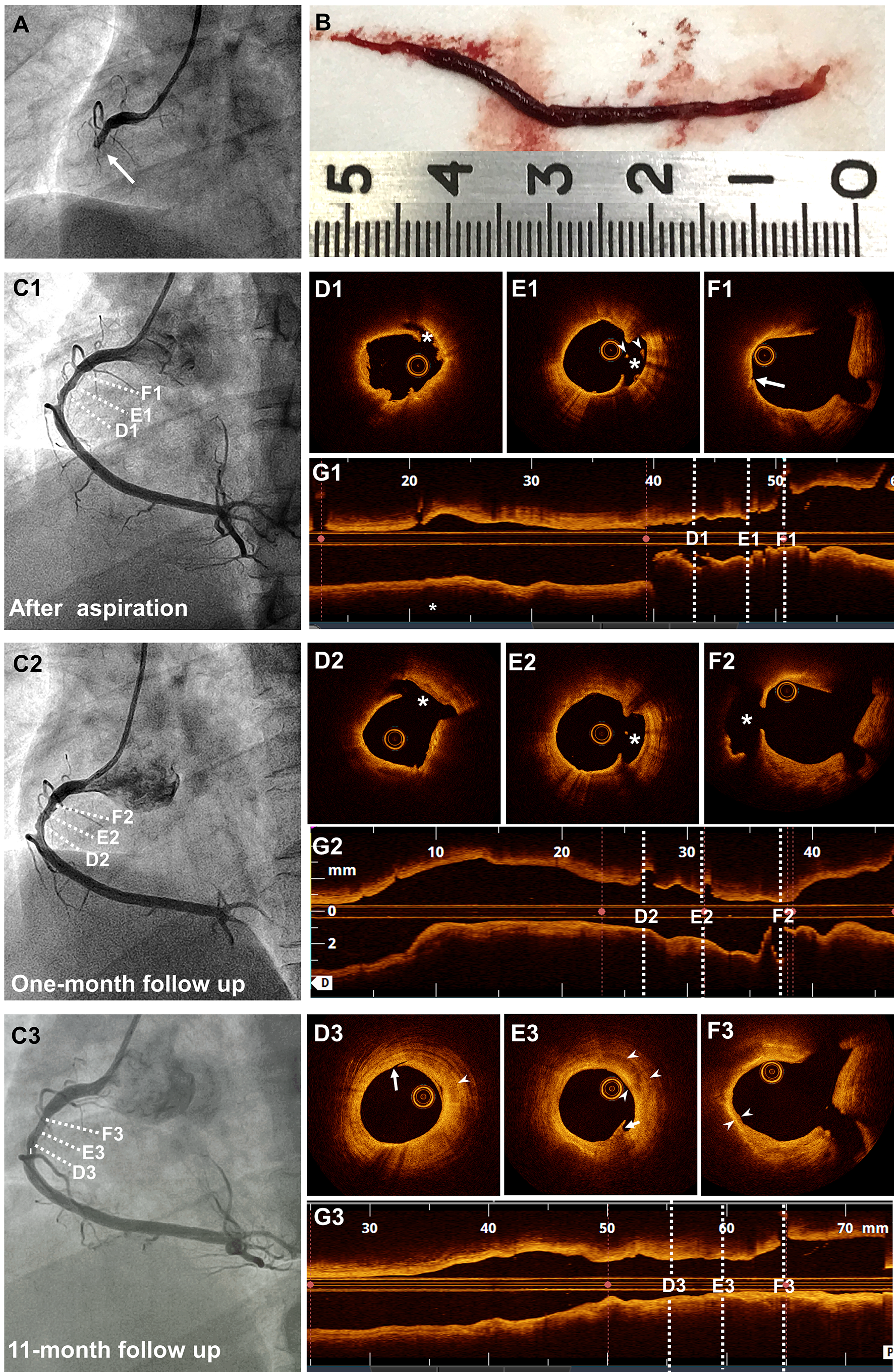
A 30-year-old man with hypertension presented to the emergency department with a 30-minute episode of diaphoresis and chest pain. An acute inferior myocardial infarction (MI) was diagnosed based on an electrocardiogram. An emergent coronary angiography showed total occlusion in the mid-right coronary artery (RCA) (Figure A). A giant thrombus was withdrawn using an aspiration catheter (Figure B), and the RCA flow was completely restored (Figure C1). Optical coherence tomography (OCT) revealed 2 plaque ruptures in the culprit lesion and a vulnerable plaque fissure in the non-culprit lesion (Figure D1-G1; Video 1). No further procedures were done. At one-month follow-up, coronary angiography (Figure C2) and OCT demonstrated unchanged plaque ruptures in the culprit lesion and progression from plaque fissure to rupture in the non-culprit lesion (Figure D2-G2; Video 2). Coronary angiography (Figure C3) and OCT at the 11-month follow-up detected resolution of plaque ruptures with layered pattern, suggestive of a healing process (Figure D3-G3; Video 3).
Our case describes OCT-based thrombus aspiration alone in the management of plaque rupture with thrombus at the RCA. Serial OCT imaging provides compelling evidence of multiple plaque ruptures healing without stenosis progression, which may be related to aggressive lowering of low-density lipoprotein cholesterol (from 3.0 mmol/L to 0.5 mmol/L). Another observation was that it is important to ensure the removal of the guidewire before conducting the OCT pullback in order to avoid wire artifacts.
Affiliations and Disclosures
From the Division of Cardiology, Beijing Luhe Hospital, Capital Medical University, Beijing, China.
Disclosures: The authors report no financial relationships or conflicts of interest regarding the content herein.
Address for correspondence: Jincheng Guo, MD, Division of Cardiology, Beijing Luhe Hospital, Capital Medical University, Tongzhou District, Beijing 101149, China. Email: guojcmd@126.com


