Cardiac Lymphoma as a Cause of Dyspnea in an Immunosuppressed Patient: The Importance of the Endomyocardial Biopsy
© 2024 HMP Global. All Rights Reserved.
Any views and opinions expressed are those of the author(s) and/or participants and do not necessarily reflect the views, policy, or position of the Journal of Invasive Cardiology or HMP Global, their employees, and affiliates.
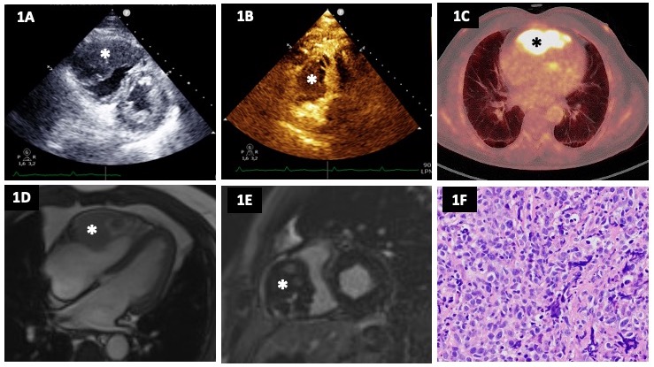
A 69-year-old man who underwent a liver transplant in 2014 due to hepatitis C virus hepatopathy was evaluated for dyspnea on exertion and chest pain. A stress echocardiogram was requested, revealing a heterogeneous mass with poorly defined borders infiltrating the right ventricle (RV) (Figure A). Due to suspicion of malignancy, the study was complemented with an echocardiographic contrast agent, which showed a heterogeneous uptake of the mass that confirmed its vascularization (Figure B; Video). The patient was admitted to the hospital for further study.
In the computed tomography (CT) body scan, no evidence of another primary tumor or distant metastasis was found. The positron emission tomography (PET)-CT showed pathological uptake at the heart (maximum standardized update value: 18.08) (Figure C).
Magnetic resonance imaging was also performed, which confirmed that the tumor infiltrated the anterior and lateral walls of the RV, had heterogeneous contours, and measured 6 x 4 x 7 cm (Figure D and E). The mass caused partial occlusion of the RV cavity without affecting the atrioventricular groove or the right coronary ostium. It showed intermediate intensity in the T2 and short-tau inversion recovery sequences, heterogeneous perfusion in perfusion sequences, and diffuse uptake in late gadolinium enhancement.
Finally, the endomyocardial biopsy allowed us to obtain a definitive diagnosis of post-transplant diffuse germinal center B-lymphoproliferative tumor located in the heart (Figure F). The patient is currently being treated with chemotherapy with a positive response, as confirmed by the follow-up PET-CT.
Primary cardiac lymphoma is an extremely rare tumor, representing less than 2% of primary cardiac tumors. Its prognosis is poor, although early diagnosis and appropriate chemotherapy treatment can enhance survival rates. It requires a high level of suspicion, especially in immunosuppressed patients, as it is the only risk factor described in the literature.1-3 In this case, the use of an echocardiographic contrast agent initially raised suspicion, and the multimodal imaging study along with the endomyocardial biopsy subsequently confirmed the diagnosis.
Affiliations and Disclosures
From the 1Department of Cardiology, La Paz University Hospital, Madrid, Spain; 2Facultad de Ciencias Biomédicas y de la Salud, Departamento de Medicina, Universidad Europea de Madrid, Madrid, Spain; 3Hospital Universitario La Paz, IdiPaz Research Institute, Madrid, Spain.
Disclosures: The authors report no financial relationships or conflicts of interest regarding the content herein.
Address for correspondence: Lucía Fernández Gassó, MD, PhD, P.º de la Castellana, 261, Fuencarral-El Pardo, Madrid 28046, Spain. Email: lucynandezga@gmail.com; X: @LuciaFGasso
References
- Asleh R, Alnsasra H, Habermann TM, Briasoulis A, Kushwaha SS. Post-transplant lymphoproliferative disorder following cardiac transplantation. Front Cardiovasc Med. 2022;9:787975. doi: 10.3389/fcvm.2022.787975
- Miguel CE, Bestetti RB. Primary cardiac lymphoma. Int J Cardiol. 2011;149(3):358-63. doi: 10.1016/j.ijcard.2010.02.016
- Gralewski J, Babu D. Diffuse large b-cell lymphoma associated with chronic inflammation and fibrin-associated large b-cell lymphoma. In: Crane GM, Loghavi S, eds. Precision Molecular Pathology of Aggressive B-Cell Lymphomas. Springer; 2024:339-350.