

Thebesian Veins Causing Severe Myocardial Ischemia Due to Coronary Steal
© 2025 HMP Global. All Rights Reserved.
Any views and opinions expressed are those of the author(s) and/or participants and do not necessarily reflect the views, policy, or position of the Journal of Invasive Cardiology or HMP Global, their employees, and affiliates.
We present a case of prominent Thebesian veins arising from the diagonal branch of the left anterior descending coronary artery (LAD), resulting in severe myocardial ischemia in the LAD territory. In the vast majority of cases, Thebesian veins are a silent finding; however, in this case, they had clinically important implications.
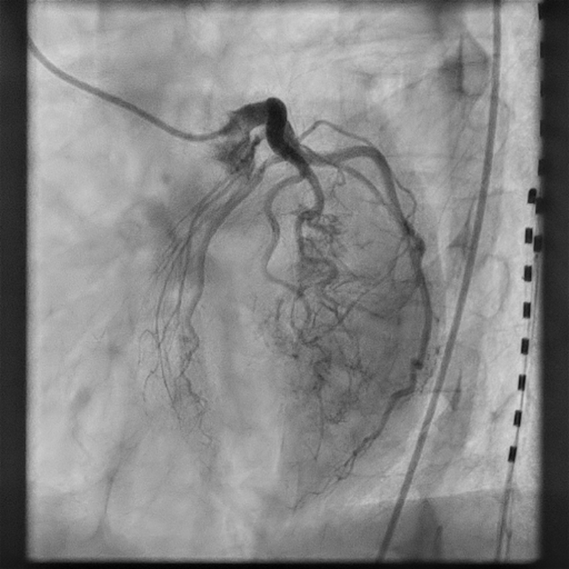
A 55-year-old woman with a history of hypothyroidism, chronic obstructive pulmonary disease, tobacco use, and anorexia nervosa was seen in the cardiology clinic for exertional chest pain and shortness of breath. A dobutamine stress test was recommended to evaluate symptoms of chest pain and shortness of breath. The stress echocardiogram was markedly abnormal, showing severe hypokinesis to akinesis in the mid-to-distal LAD territory with stress-induced decrease in ejection fraction (Videos 1 and 2). Therefore, she was referred for cardiac catheterization. On cardiac catheterization, she was found to have widely patent epicardial arteries with prominent Thebesian veins arising from a diagonal branch. There was significant shunting of the blood via a diagonal branch directly into the left ventricular cavity (Figure; Video 3).
Thebesian veins are valveless vascular channels that provide a direct connection between the coronary arteries and the cardiac chambers. Thebesian and bronchial veins are examples of physiological shunts. These vascular channels in the heart were first described by anatomist Adam Christian Thebesius in 1708 and have no clear function.1 It is uncommon to see these channels on a coronary angiogram, and their prevalence is suggested to be around 0.1%.2 It is very rare for Thebesian veins to cause myocardial ischemia. It is important to recognize these channels when they do cause ischemia, as in the current case. Currently, there is no clear clinical guidance to guide treatment if they are the suspected etiology of myocardial ischemia.
Affiliations and Disclosures
Gurpreet Singh, MD1,2; Vien Le, MD2; Vishnu Patlolla, MD2
From the 1Department of Cardiology, Mayo Clinic, Rochester, Minnesota; 2Department of Cardiology, Mayo Clinic Health System, Eau Claire, Wisconsin.
Disclosures: The authors report no financial relationships or conflicts of interest regarding the content herein.
Consent statement: Patient permission and consent were obtained for publication of this case report/imaging case.
Artificial intelligence (AI) statement: No generative AI or AI-assisted technologies were used to prepare this article.
Address for correspondence: Gurpreet Singh, MD, Department of Cardiology, 1221 Whipple St, Eau Claire, WI 54703, USA. Email: singh.gurpreet@mayo.edu
References
- Ansari A. Anatomy and clinical significance of ventricular Thebesian veins. Clin Anat. 2001;14(2):102-10. doi:10.1002/1098-2353(200103)14:2<102::AID-CA1018>3.0.CO;2-4
- Carlyle A, Pitney M. Is Thebesian venous drainage a part of a clinical syndrome? Heart Lung Circ. 2013;22(S1):S140-S141. doi:10.1016/j.hlc.2013.05.335


