Video Supplement: Transapical Device Closure of a Submitral Left Ventricular Pseudoaneurysm After Mitral Valve Replacement
Video Supplement to "Transapical Device Closure of a Submitral Left Ventricular Pseudoaneurysm After Mitral Valve Replacement" (Clinical Image).
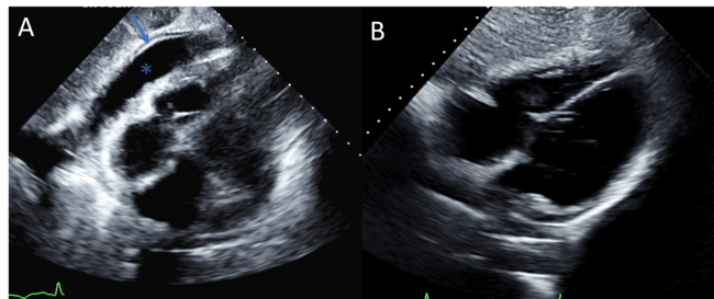
Video 1. TTE in parasternal long-axis view demonstrating the submitral left ventricular pseudoaneurysm, with color Doppler confirming flow across the pseudoaneurysm neck. TTE = transthoracic echocardiography.
Video 2. Fluoroscopic image showing simultaneous left CAG during transapical puncture with a micropuncture needle, used to delineate the left anterior descending artery and avoid coronary injury. CAG = coronary angiography; CAU = caudal; PDA = patent ductus arteriosus; RAO = right anterior oblique.
Video 3. Combined transesophageal echocardiographic and fluoroscopic imaging showing the catheter advanced across the pseudoaneurysm neck and positioned within the pseudoaneurysm cavity before device deployment. CRA = cranial; LAO = left anterior oblique; PDA = patent ductus arteriosus.
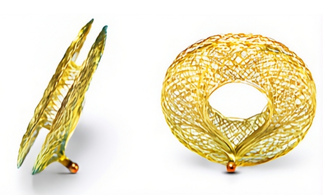
Video 4. Combined transesophageal echocardiographic and fluoroscopic imaging showing release of the muscular ventricular septal defect occluder after confirmation of stable position across the pseudoaneurysm neck. CRA = cranial; LAO = left anterior oblique; patent ductus arteriosus.
Video 5. Final left ventricular angiography in left anterior oblique cranial projection showing both occluder devices in position, with no residual opacification of the pseudoaneurysm or access-site leak. CRA = cranial; PDA = patent ductus arteriosus; RAO = right anterior oblique.
© 2026 HMP Global. All Rights Reserved.
Any views and opinions expressed are those of the author(s) and/or participants and do not necessarily reflect the views, policy, or position of the Journal of Invasive Cardiology or HMP Global, their employees, and affiliates.