9.2 Combined Imaging/Physiology Catheters: Devices Everyone Should Have and Use and How AI will Make Them Even Better
Problem Presenter: Jimmy Kerrigan
Problem Presenter: Jimmy Kerrigan
These proceedings summarize the educational activity of the 18th Biennial Meeting of the International Andreas Gruentzig Society held January 27 to 30, 2026, in Puerto Ayora, Santa Cruz, Ecuador.
Faculty Disclosures Sponsors
2026 IAGS Summary Document
Statement of problem or issue
Intravascular imaging (IVI) used in percutaneous coronary intervention (PCI) saves lives. A large network meta-analysis of 22 trials with over 15,000 patients and weighted mean duration of follow-up of 25 months (range 6-60 months) demonstrated this (Figure 1).[1]

Similarly, the 10-year results of the FAME trial demonstrated that physiology-guided PCI using fractional flow reserve (FFR) measured with intracoronary pressure wires reduced subsequent events over this very long interval.[2]
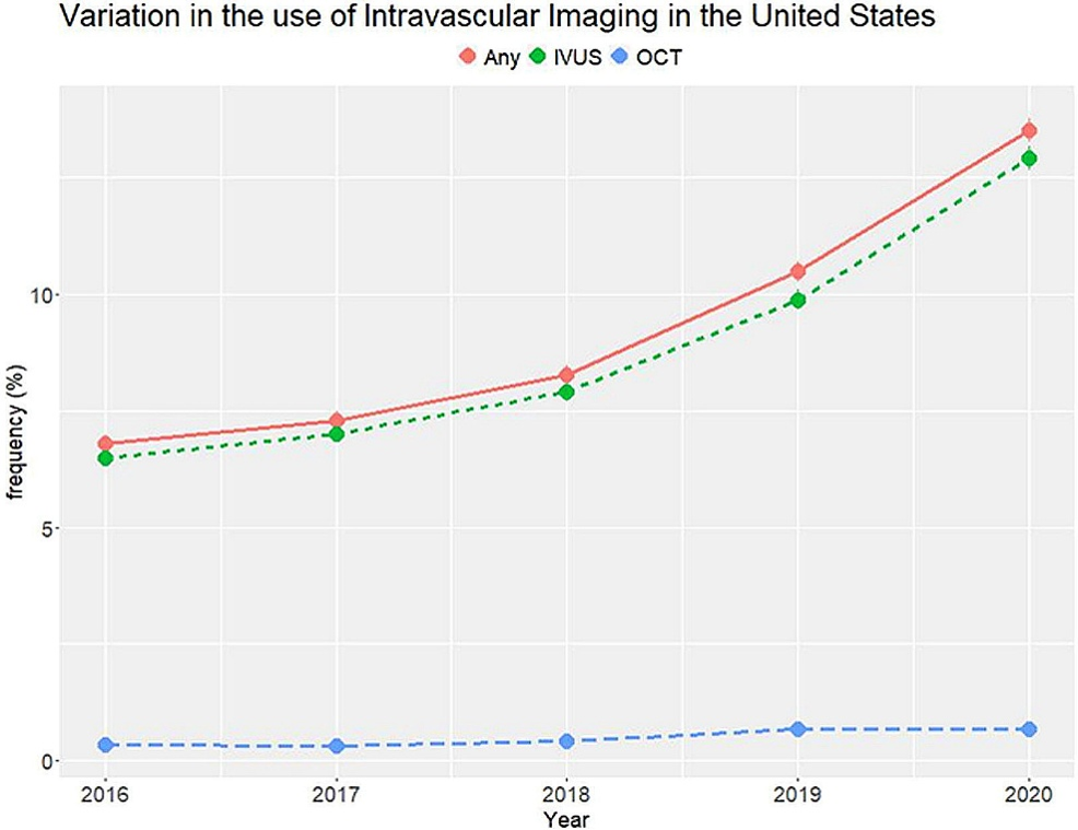
Despite these findings, use of either IVI or coronary physiology in the United States remains low (Figure 2).[3,4] Although there has been a slight trend of increased use over the past decade, overall use of IVI is approximately 15% of PCI cases. [3] However, recent reports from the National Cardiovascular Data Registry (NCDR) and other surveys suggest the use of coronary physiology is approaching 30% to 35% of cases.[4]
Gaps in current knowledge
The largest knowledge gaps are:
- How to increase the use of IVI and coronary physiology by operators.
- How to get adequate payments/reimbursements for these procedures.
- How to get hospitals to invest in the appropriate equipment.
- Will alternative methods like angio-derived physiology measures be useful?
- Will combinations of IVI and physiology be superior to either alone?
Possible solutions or future directions
Quantitative angiographic analysis systems incorporating computational fluid dynamics equations can estimate several parameters from angiograms that previously could be determined only by measuring intracoronary pressures directly with a pressure-sensing wire. Indices like FFR, iFR, and others can now be estimated by angiography. The advantages of these angiography-based systems are that angiographic imaging is the standard imaging modality by which coronary arteries are evaluated and interventional procedures are conducted. The disadvantages are that processing time for angiography-based analyses can be long, and images must be clear and uncomplicated, without convolutions, tortuosity, and overlap. Catheter technology also is evolving, and it is possible that catheters incorporating both imaging and physiology will become available. Whether these newer methodologies will replace previous ones is still undetermined. Issues to be addressed include workflow integration, operator and staff familiarity and comfort, and demonstrating that use of the derived information influences and improves patient care and is not done just for the sake of using catheters. Costs, facility reimbursement (or, penalties for non-use), and value will also be important considerations influencing utilzation.[5,6]
References
- Stone GW, et al. Intravascular imaging-guided coronary drug-eluting stent implantation: an updated network meta-analysis. Lancet. 2024 Mar 2;403(10429):824-837. doi: 10.1016/S0140-6736(23)02454-6. Epub 2024 Feb 21. PMID: 38401549.
- Collet C, et al. Fractional flow reserve-guided percutaneous coronary intervention versus medical therapy for stable coronary artery disease: long-term results of the FAME 2 trial. Nat Med. 2026 Jan;32(1):318-324. doi: 10.1038/s41591-025-04132-5. Epub 2026 Jan 15. PMID: 41540107.
- Chaturvedi A, et al. Regional Disparities and Predictors of Intracoronary Imaging Use During Percutaneous Coronary Intervention in the United States. Am J Cardiol. 2025 Jul 3;255:1-9. doi: 10.1016/j.amjcard.2025.06.017. Epub ahead of print. PMID: 40617275.
- Carvalho PEP, et al. Contemporary coronary physiology practice: An international survey of interventional cardiologists. Cardiovasc Revasc Med. 2026 Mar 29:S1553-8389(26)00056-4. doi: 10.1016/j.carrev.2026.02.011. Epub ahead of print. PMID: 41912370.
- Volleberg RHJA, et al. Combining Optical Coherence Tomography and Fractional Flow Reserve for Decision Making in Percutaneous Coronary Intervention: Insights From the FUSION Study. J Soc Cardiovasc Angiogr Interv. 2025 Oct 7;4(11):104001. doi: 10.1016/j.jscai.2025.104001. PMID: 41324056.
- Fearon WF, et al; ALL-RISE Investigators. Angiography-Derived Fractional Flow Reserve to Guide PCI. N Engl J Med. 2026 Mar 29. doi: 10.1056/NEJMoa2600949. Epub ahead of print. PMID: 41910384.
© 2026 HMP Global. All Rights Reserved.
Any views and opinions expressed are those of the author(s) and/or participants and do not necessarily reflect the views, policy, or position of the Journal of Invasive Cardiology or HMP Global, their employees, and affiliates.