

Alpha-Loop Snaring Technique: Facilitated Snaring Technique for Fully-Deployed and Deformed Coronary Stent Protruding Into the Aorta
Keywords: snaring, stent, coronary, deformed stent, angioplasty, PCI, IVUS
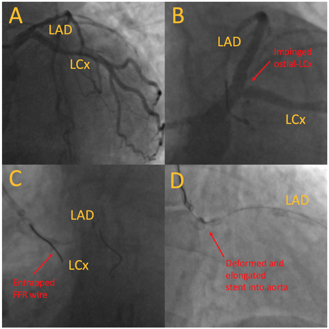
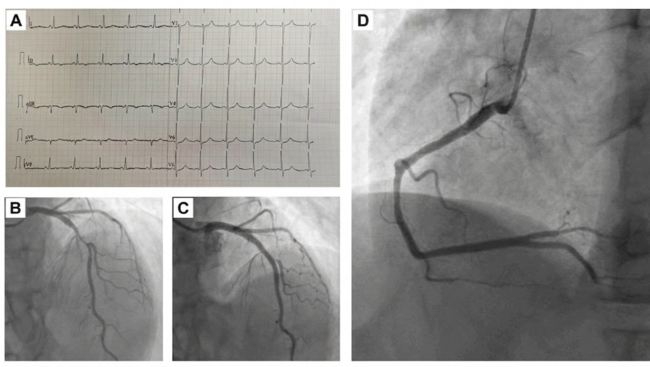
A 59-year-old female was admitted for non-ST-elevation myocardial infarction (NSTEMI) and Killip class 3 heart failure. Echocardiogram showed left ventricular ejection fraction (LVEF) of 30%. Coronary angiogram showed moderate to severe stenosis over the ostial-proximal left anterior descending artery (LAD) (Figure 1A), with minor disease over the left circumflex artery (LCx) and right coronary artery (RCA). Fractional flow reserve (FFR) and instantaneous wave-free ratio (iFR) performed across the LAD lesion were 0.78 and 0.86, respectively. The FFR wire was then parked at the LCx for side branch protection. Intravascular ultrasound (IVUS) showed significant disease over the distal left main (LM) to the mid-LAD. IVUS-guided provisional stenting of the LM-LAD was performed with 2 overlapping drug-eluting stents. The final angiogram showed an impinged ostial LCx (Figure 1B), used for functional study. The jailed FFR wire at the LCx removed and attempted to recross the pinched LCx, but was trapped at the proximal LCx, unable to be advanced or pulled (Figure 1C). The wire was removed with difficulty after numerous attempts, with microcatheter and coronary balloon assistance. However, it resulted in severely deformed and elongated stents, with long segment protrusion into the aorta (Figure 1D). “Ping-pong” guiding with a 9-Fr JL4 via right femoral artery was performed. Distorted stents were wired in and out of stent struts intentionally with 3 guidewires via the new JL4, intending to remove the stents with a twisted wire technique, but failed (Figure 2A;Video 1).
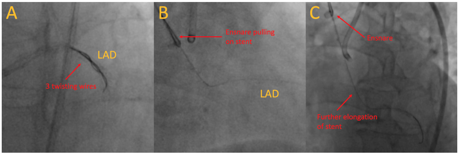
Subsequently, an EN Snare endovascular snare system (Merit Medical) was sent along the original true-lumen guidewire to pull the stents out. Initially, the protruding stent, along with the remaining stents inside the coronary artery, were moving out (Figure 2B; Video 2). However, with further pulling, the middle part of the stent weakened and further elongated to the verge of breaking (Figure 2C). With further pulling, the stent would break, risking both stent embolization and long stent protrusion inside the aorta.
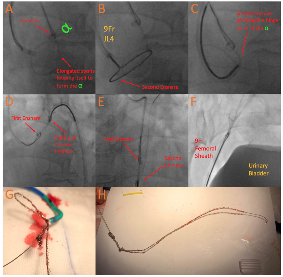
We came up with the alpha-loop snaring technique. We pushed the system forward, so that the weakened mid stent formed an alpha-shaped loop (Figure 3A). Another EN Snare was then sent via the 9-Fr JL4 guiding catheter, snaring the hinge point of the “alpha” (Figures 3B and 3C; Video 3), bypassing the weakened point of the elongated stent. All the remaining parts of the stents were successfully pulled with the second Ensnare (Figure 3D; Video 4). With both Ensnare tightened, the stents were sent to the lower descending aorta (Figure 3E; Video 5), where the first snare was released. The stents were externalized (Figures 3F-3H; Video 6). The patient remained hemodynamically stable throughout the procedure. IVUS-guided LM-bifurcation stenting with Culotte technique was performed with optimal result (Video 7).
To our knowledge, this is the first report on our Novel approach of the alpha-loop snaring technique, which can salvage a failing snaring attempt of a completely deployed and dislodging coronary stent.
Affiliations and Disclosures
From the Cardiology Division, Department of Medicine, Queen Elizabeth Hospital, Hong Kong.
Disclosure: The authors have completed and returned the ICMJE Form for Disclosure of Potential Conflicts of Interest. The authors report no conflicts of interest regarding the content herein.
The authors report that patient consent was provided for publication of the images used herein.
Manuscript accepted March 25, 2022.
Address for correspondence: Michael Chiang, MBBS, Cardiology Division, Department of Medicine, Queen Elizabeth Hospital, 30 Gascoigne Road, Kowloon, Hong Kong. Email: michaelcschiang@gmail.com


