

Catheter-Induced Left Ventricular Perforation and Cardiac Tamponade During Left Heart Catheterization
Keywords: left ventricular perforation, cardiac tamponade
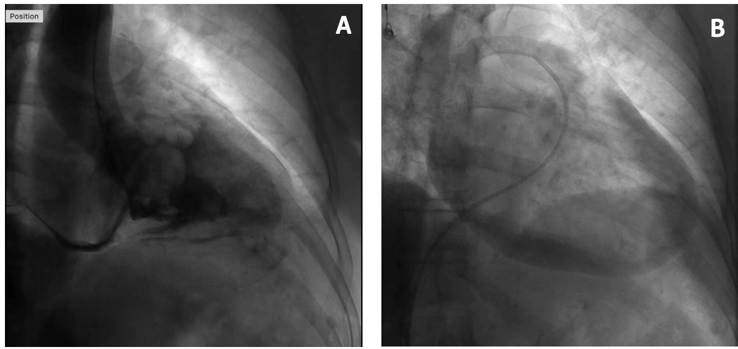
Left ventricular perforation is a rare complication of myocardial infarction, cardiac trauma, or due to an iatrogenic cause, such as cardiac catheterization, biopsy, temporary or permanent pacing, or pericardiocentesis. We present a case of a 79-year-old woman who underwent a left heart catheterization complicated by catheter-induced ventricular perforation. The aortic valve was difficult to cross with a 0.035-inch regular J-tip wire. A 5-Fr AL2 catheter and 0.035-inch regular straight wire were used instead. The AL2 catheter was then swapped with the straight pigtail catheter. The left ventriculogram showed normal left ventricular function with dye opacification into the pericardial cavity, outlining the pulmonary artery, aorta, and left atrium (Figure 1; Video Series), consistent with intraprocedural ventricular perforation with consequent hemorrhagic pericardial effusion (Figure 2; Video Series). The patient was hemodynamically stable initially, however, ~45 minutes post procedure, her systolic blood pressure reduced to 50 mm Hg. Right heart catheterization revealed cardiac tamponade physiology. Emergent pericardiocentesis was performed and 400 mL of frank blood drained. The patient was stabilized and sent to a cardiothoracic center for pericardial patch repair for the probable perforation. She remained stable and contrast echocardiogram confirmed the absence of persistent communication between the left ventricle and the pericardium.
Iatrogenic ventricular perforation of the myocardial wall is a rare but life-threatening complication. It has been described using pulmonary artery catheter, pigtail catheter, and Judkins catheter.1-3 Straight wire and catheters can be used to cross the aortic valve for left ventriculogram, however the risk of perforation is higher compared to a J-tip wire. Prompt recognition of cardiac tamponade and pericardial drain insertion is vital, but surgical patch repair may be required for definitive treatment. This case highlights the importance of increased vigilance with usage of high-risk equipment during cardiac catheterization and prompt management of cardiac tamponade.
Affiliations and Disclosures
From the Department of Cardiology, Launceston General Hospital, Launceston, Australia.
Disclosure: The authors have completed and returned the ICMJE Form for Disclosure of Potential Conflicts of Interest. The authors report no conflicts of interest regarding the content herein.
The authors report that patient consent was provided for publication of the images used herein.
Manuscript accepted May 1, 2022.
Address for correspondence: Dr Savvy Nandal, Launceston General Hospital, 274-280 Charles Street, Launceston TAS 7250, Australia. Email: savvy.nandal@gmail.com
References
1. Benito-Saz P, Garrido A, Quintana-Villamandos B, Barrio JM, Fernandez-Quero L, Hortal J. Perforation of the left ventricle wall due to the insertion of a pulmonary artery catheter. A case report. Rev Esp Anestesiol Reanim (Engl Ed). 2019;66(10):528-532. Epub 2019 Oct 4. doi:10.1016/j.redar.2019.06.001
2. Sharif H, Hussain S. Accidental left ventricular perforation during coronary angiogram. J Coll Physicians Surg Pak. 2014;24(Suppl 2):S74-S75.
3. Provaznik Z, Holzamer A, Camboni D, et al. Perforation of myocardial wall and great vessels after cardiovascular interventions-a 5-year analysis. J Thorac Dis. 2017;9(12):5288-5294. doi:10.21037/jtd.2017.10.11


