





Evolving Tools for Residual Disease Detection in Lymphoma
In the management of diffuse large B-cell lymphoma (DLBCL), achieving a complete response at the end of treatment is the goal. Yet relapse remains a persistent challenge, with 30%–40% of patients ultimately experiencing disease recurrence.1 This underscores a critical gap in how we assess treatment response and residual disease, and raises an important question: are our current tools sufficient to determine who is truly in remission and who remains at risk?
PET/CT: Essential, but Not Definitive
Positron emission tomography/computed tomography (PET/CT) scans remain the standard of care for end-of-treatment (EOT) response assessment in lymphoma. By measuring metabolic activity through a radiotracer uptake, PET/CT scans provide a functional view of disease and helps categorize patients into complete metabolic response or residual disease using the Deauville scoring system.2,3
While indispensable in practice, PET/CT scans have well-recognized limitations.
-
Sensitivity is limited at low disease burden, meaning microscopic residual disease may go undetected.4
-
False positives are common, particularly in the setting of inflammation.4
-
False negatives can underestimate relapse risk.5
-
In a large dataset at 2.5 years, approximately 15% of patients with negative PET scans relapsed after end of treatment, compared with 35%–47% of PET-positive patients (Figure 1).6
What does this mean in practice? Clinicians are often left navigating ambiguity and left wondering:
-
Is residual uptake true disease or inflammation?
-
Should treatment be escalated or avoided?
While valuable, PET/CT scans alone do not provide a complete picture of residual disease and relapse risk.7
Figure 1. Progression-Free Survival in PET/CT CR-Positive and -Negative DLBCL Patients6
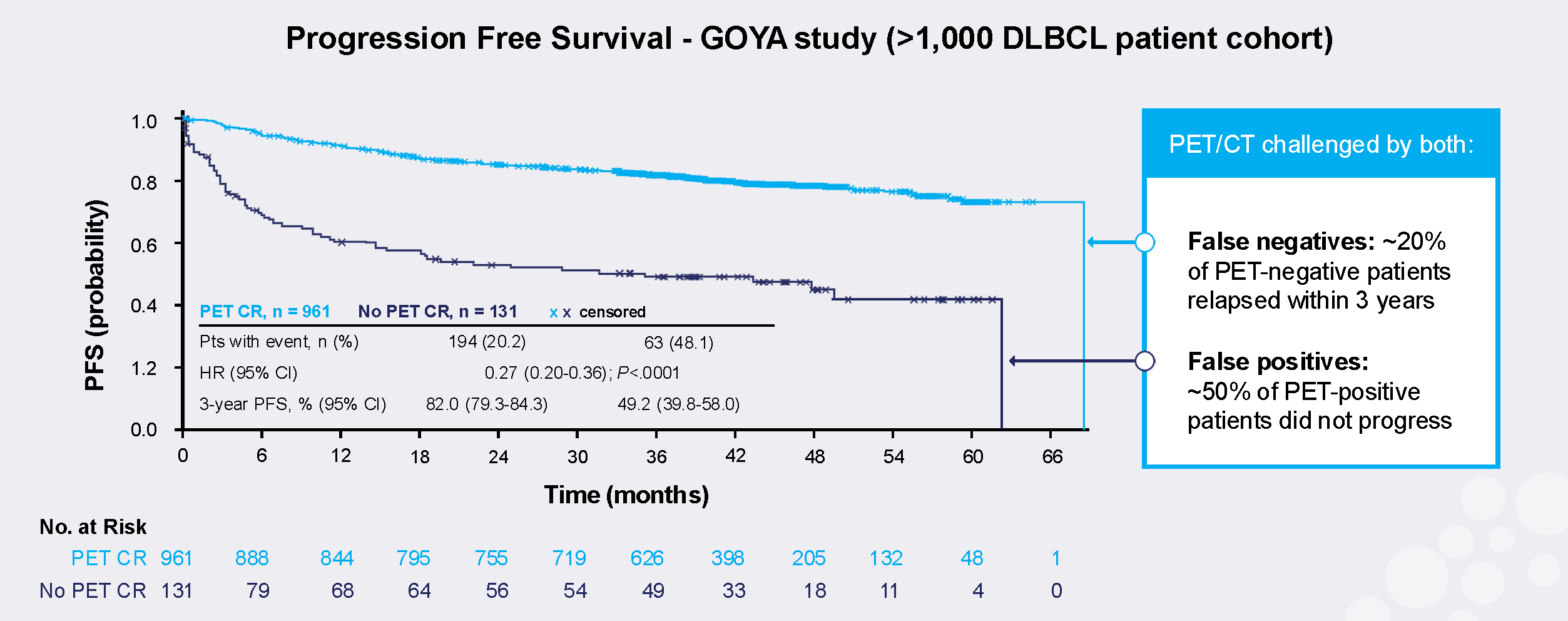
CI, confidence interval; CR, complete response; CT, computed tomography; DLBCL, diffuse large B-cell lymphoma; HR, hazard ratio; PET, positron emission tomography; PFS, progression-free survival.
Beyond Imaging: Emerging Approaches to Residual Disease Detection
Efforts to improve residual detection have included both cell-based and molecular approaches.
-
Flow cytometry can detect malignant cells but has limited sensitivity.7
-
PCR-based methods historically tracked immunoglobulin arrangements but are less scalable.7
These limitations have driven clinicians’ and researchers’ interest in more sensitive, less invasive approaches.
The Shift Toward Molecular Detection: ctDNA MRD
Over the past decade, advances in next-generation sequencing (NGS) have enabled a new approach: detecting circulating tumor DNA (ctDNA) in the bloodstream.7 Unlike imaging, which captures a static picture, ctDNA reflects tumor-derived genetic material, offering a more direct and biologically relevant measure of disease.7 Modern ctDNA-based MRD assays can identify disease at very low levels, with a sensitivity of 10–6, corresponding to about one tumor-derived DNA fragment per 1 million normal DNA fragments.7
Refining Risk Stratification: ctDNA vs PET/CT
Emerging data suggest that ctDNA MRD testing provides more precise prognostic information than PET/CT scans alone. In a study by Roschewski and colleagues, patients who were PET-negative but ctDNA-positive at EOT had significantly worse outcomes, with only 31% 2-year progression-free survival (PFS).8 In contrast, patients who were ctDNA-negative had 2-year PFS rates approaching 95%-98% (Figure 2).8
What does this mean in practice? ctDNA MRD testing can:
-
Identify patients at high risk of relapse despite a “clean” PET scan8
-
Provide reassurance in patients with deep molecular remission7
-
Help clarify ambiguous PET findings7
Figure 2: ctDNA MRD is Superior to PET/CT Imaging for Prognostication in DLBCL8
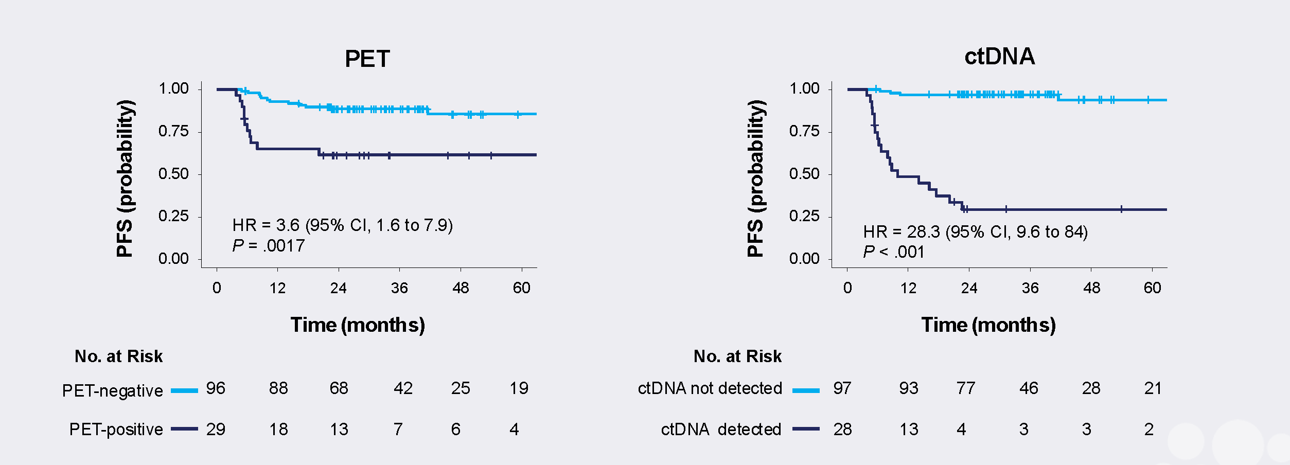
CI, confidence interval; ctDNA, circulating tumor DNA; HR, hazard ratio; PET, positron emission tomography; PFS, progression-free survival.
From Static to Dynamic Assessment
Residual disease evaluation involves both response assessment (during and at the end of treatment) and surveillance (monitoring after treatment). ctDNA supports both settings. During treatment, early molecular response may provide insight into treatment effectiveness before imaging.7 At EOT, MRD status refines the interpretation of PET/CT findings. In surveillance, serial ctDNA monitoring may detect relapse earlier than conventional methods.7 This represents a shift from a single timepoint, imaging-based model to a more dynamic, longitudinal understanding of disease biology.
Why This Matters in Practice
As treatment strategies evolve, particularly with increasing use of immunotherapies, accurate response assessment becomes even more critical. Notably, these therapies may increase false-positive PET/CT findings, further complicating interpretation.5
Integrating ctDNA MRD testing into lymphoma care may:
-
Improve risk stratification at EOT8
-
Offer opportunities for response-adapted strategies, including escalation or de-escalation of therapy7
-
Enable earlier detection of relapse, potentially allowing intervention when disease burden is lower9
As our understanding of lymphoma biology advances, so must the tools we use to assess it. ctDNA MRD testing offers a promising step toward more confident, individualized patient management.
References
1. Freeman CL, Savage KJ, Villa DR, et al. Long-term results of PET-guided radiation in patients with advanced-stage diffuse large B-cell lymphoma treated with R-CHOP. Blood. 2021;137(7):929-938. doi:10.1182/blood.2020005846
2. Cheson BD, Fisher RI, Barrington SF, et al. Recommendations for initial evaluation, staging, and response assessment of Hodgkin and non-Hodgkin lymphoma: the Lugano classification. J Clin Oncol. 2014;32(27):3059-3068. doi:10.1200/JCO.2013.54.8800
3. Al Tabaa Y, Bailly C, Kanoun S. FDG-PET/CT in lymphoma: where do we go now? Cancers (Basel). 2021;13(20):5222. doi:10.3390/cancers13205222
4. Cronin CG, Swords R, Truong MT, et al. Clinical utility of PET/CT in lymphoma. AJR Am J Roentgenol. 2010;194(1):W91-W103. doi:10.2214/AJR.09.2637
5. Kasamon YL, Jones RJ, Wahl RL. Integrating PET and PET/CT into the risk-adapted therapy of lymphoma. J Nucl Med. 2007;48:19S-27S. doi:10.2967/jnumed.106.037556
6. Kostakoglu L, Martelli M, Sehn LH, et al. End-of-treatment PET/CT predicts PFS and OS in DLBCL after first-line treatment: results from GOYA. Blood Adv. 2021;5(5):1283-1292. doi:10.1182/bloodadvances.2020002690
7. Zhang S, Wang X, Yang Z, et al. Minimal residual disease detection in lymphoma: methods, procedures and clinical significance. Front Immunol. 2024;15:1430070. doi:10.3389/fimmu.2024.1430070
8. Roschewski M, Kurtz DM, Westin JR, et al. Remission assessment by circulating tumor DNA in large B-cell lymphoma. J Clin Oncol. 2025;43(34):3652-3661. doi:10.1200/JCO-25-01534
9. Herrera AF, Armand P. Minimal residual disease assessment in lymphoma: methods and applications. J Clin Oncol. 2017;35(34):3877-3887. doi:10.1200/JCO.2017.74.5281
© 2026 HMP Global. All Rights Reserved.