


DLL3 Testing and Scoring: Practical Considerations for Pathologists
Dr Anja Roden examines practical considerations for delta-like ligand 3 (DLL3) testing and interpretation, including why immunohistochemistry protein expression assessment differs from next-generation sequencing-based genomic testing. This video addresses specimen adequacy, staining interpretation, scoring, and the important distinction between “DLL3-positive” and “DLL3-high,” emphasizing clear, assay-specific reporting as testing frameworks evolve.
Transcript
Anja Roden, MD: I'm Anja Roden. I'm one of the thoracic and surgical pathologists at Mayo Clinic in Rochester, Minnesota. I'm very interested in biomarkers. I have been the immunostains lab director for many years and was involved in the validation and testing of many of the predictive biomarkers.
So for the treatment with anti-DLL3, we have to remember that the anti-DLL3, which is an antibody, will link to the antigen that is on the cell surface. And therefore, we need to look whether this antigen, which is a protein, is actually present on the cell surface. And this may not be linked to any mutation or any other molecular alterations. There are various mechanisms why an antibody, an antigen, a protein, may be expressed on the cell surface or not.
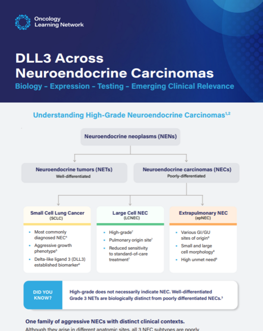
So to best test whether the antigen is present on the cell surface, immunohistochemistry using an antibody that is targeting DLL3 is the best method. Furthermore, I'm not aware of any studies that have looked into DLL3 molecular alterations and have those linked to anti-DLL3 treatment. And to my knowledge, many of the large NGS panels don't even have the DLL3 gene in them.
So ideally, we want to have a specimen that is formalin-fixed, paraffin-embedded, and is not decalcified because decalcification can interfere with the immunohistochemistry. And you want to have a validated antibody, of course. You need to follow the CAP guidelines. This is a predictive biomarker, so we need to test at least 20 positive and 20 negative samples. In our lab, we usually do way more than that, but the CAP guidelines are 20 positive and 20 negative cases.
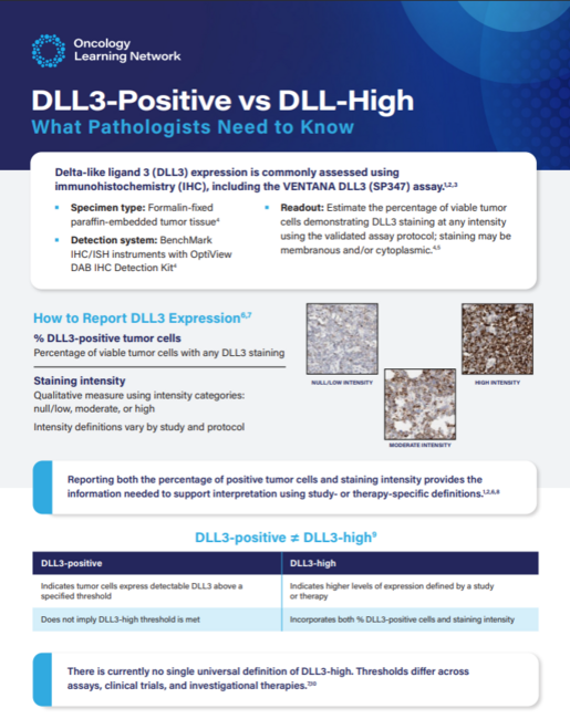
Once you have validated the antibody, you then can use it, and you look for the percentage of positive tumor cells. This is a cytoplasmic and membranous expression. Now, in small cell carcinomas, whether it's cytoplasmic or membranous is often difficult to distinguish, but the general rule is cytoplasmic and membranous expression. And we do report the percentage of tumor cell expression. However, to my knowledge, there are various trials out there that use various percentages of tumor cell expression for inclusion criteria.
So we can't really report positive or negative unless you only report it for a very specific trial for which you know which percentage of tumor cell positivity the trial is including for. Otherwise, you would just report the percentage of DLL3-positive tumor cells. We also have to report which clone of the immunostain we use and that it is on formalin-fixed, paraffin-embedded tissue.
Various clinical trials have various inclusion criteria. If you say DLL3 is positive based on—and I'm just making that up—a clinical trial that says if more than 25% of tumor cells express DLL3, we will include the patient, then if you have a tumor that expresses 50% of the tumor cells express DLL3, you will call this positive.
However, there are other clinical trials that not only include the percentage of tumor cells, but also the intensity of expression. So meaning no expression, weak expression, moderate expression, and strong expression: 0, 1+, 2+, and 3+. And so in some of these clinical trials, the expression has to be—the expression intensity has to be 3+. And then you would call this high expression if it's an intense, strongly intense expression pattern. So you can't interchange highly expressing from this positive. This cannot be interchanged.
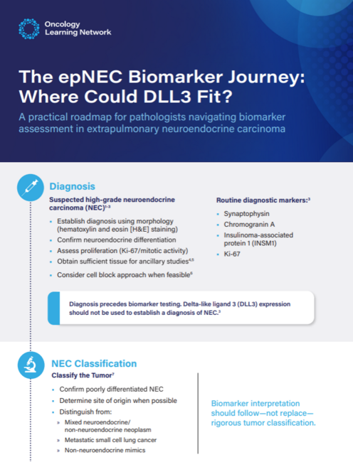
Of course, the specimen should be included. The site of the biopsy or the resection, we need to include the block number, so like A1, A2, B1, B2, so that we always know which block was used. Then we need to include the name of the antibody, which would be DLL3, the clone of the antibody, and the company from which we have the antibody purchased. And then under the results section or the interpretation section, we will say like 50% of tumor cells showing cytoplasmic and membranous staining.
Various labs use various comments, but the comment needs to include that this was performed on formalin-fixed, paraffin-embedded tissue and that if the test was performed on decalcified tissue, the results may not reflect the real staining.
So in general, the DLL3 antibody or DLL3 testing is an immunostain. It will report the percentage of DLL3-positive tumor cells. This is needed for a clinical trial, and there are certain requirements to include patients into that clinical trial. The trial clinician should talk with the pathologist so that the pathologist can make sure that all the important information for that trial is included in the report.
 Anja C. Roden, MD, is Professor of Laboratory Medicine and Pathology and is a board-certified anatomic and clinical pathologist with special interest in thoracic pathology.
Dr Roden received her medical training at the Humboldt University in Berlin and Technical University in Dresden, Germany. After training in general surgery and working in basic immunology science, she completed her anatomic and clinical pathology residency, surgical pathology fellowship, and pulmonary Mayo Clinic scholarship at Mayo Clinic in Rochester, US. Subsequently, she joined the staff of the Department of Laboratory Medicine and Pathology at Mayo Clinic in Rochester. Dr Roden also serves as Pulmonary Pathology Fellowship director and head of the thoracic transplant section.
© 2026 HMP Global. All Rights Reserved.
Any views and opinions expressed are those of the author(s) and/or participants and do not necessarily reflect the views, policy, or position of Oncology Learning Network or HMP Global, their employees, and affiliates.